Agemura Tomoya, Hasegawa Tetsuo, Yari Shinya, Kikuta Junichi, Ishii Masaru
Department of Immunology and Cell Biology, Graduate School of Medicine and Frontier Biosciences, Osaka University, 2-2 Yamada-oka, Suita, Osaka, 565-0871, Japan.
WPI-Immunology Frontier Research Center, Osaka University, 2-2 Yamada-oka, Suita, Osaka, 565-0871, Japan.
Inflamm Regen. 2022 Jun 2;42(1):17. doi: 10.1186/s41232-022-00206-w.
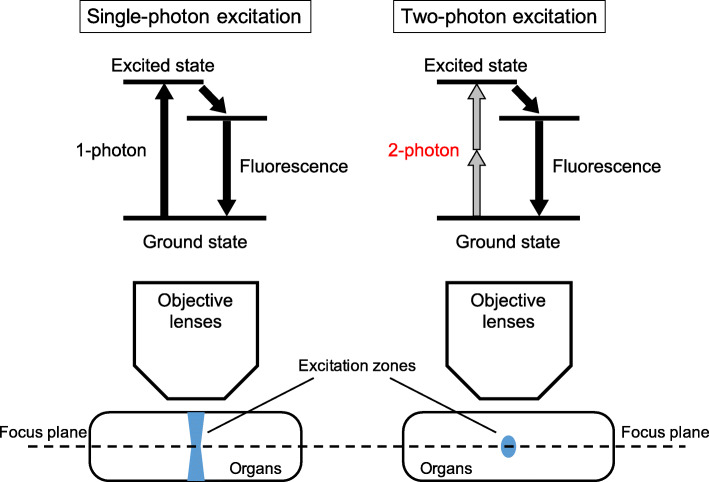
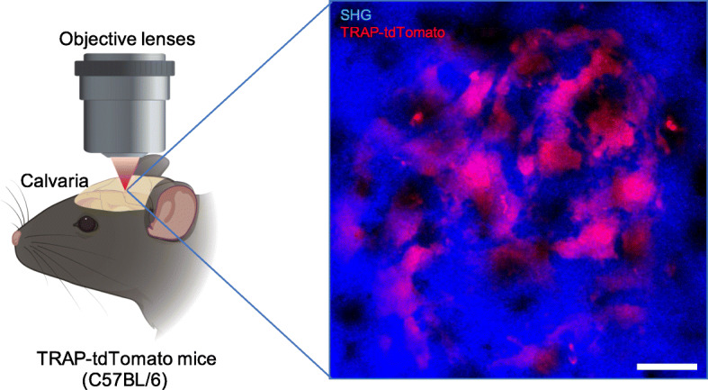
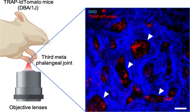
Osteoclasts are myeloid lineage cells with a unique bone-destroying ability that maintains bone homeostasis together with bone formation by osteoblasts. An advanced intravital imaging system using a two-photon microscopy has enabled the observation and evaluation of osteoclast dynamics and behaviors in the bone marrow of living mice. Using this system, it has become clear that pathological osteoclasts under inflamed conditions differ from physiological osteoclasts under a steady-state. Recently, we identified novel osteoclast precursors in arthritis, called arthritis-associated osteoclastogenic macrophages (AtoMs), which differentiate into pathological osteoclasts and induce inflammatory bone destruction. In this review, we introduce the in vivo imaging of physiological and pathological osteoclasts and their differentiation mechanism.
破骨细胞是具有独特骨破坏能力的髓系谱系细胞,它与成骨细胞的骨形成一起维持骨稳态。一种使用双光子显微镜的先进活体成像系统能够观察和评估活小鼠骨髓中破骨细胞的动态和行为。利用该系统,已经明确炎症条件下的病理性破骨细胞与稳态下的生理性破骨细胞不同。最近,我们在关节炎中鉴定出了新的破骨细胞前体,称为关节炎相关破骨细胞生成巨噬细胞(AtoMs),它们分化为病理性破骨细胞并诱导炎性骨破坏。在这篇综述中,我们介绍生理性和病理性破骨细胞的体内成像及其分化机制。