Avesani Martina, Kang Sok-Leng, Jalal Zakaria, Thambo Jean-Benoit, Iriart Xavier
Department of Pediatric and Congenital Cardiology, M3C National Reference Centre, Bordeaux University Hospital, Bordeaux, France.
Department of Pediatric Cardiology, Alder Hey Children's Hospital, Liverpool, United Kingdom.
Front Pediatr. 2022 May 19;10:894472. doi: 10.3389/fped.2022.894472. eCollection 2022.
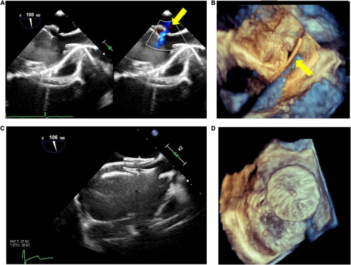
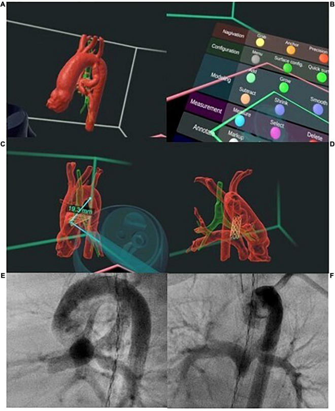
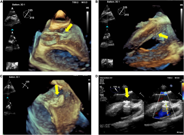
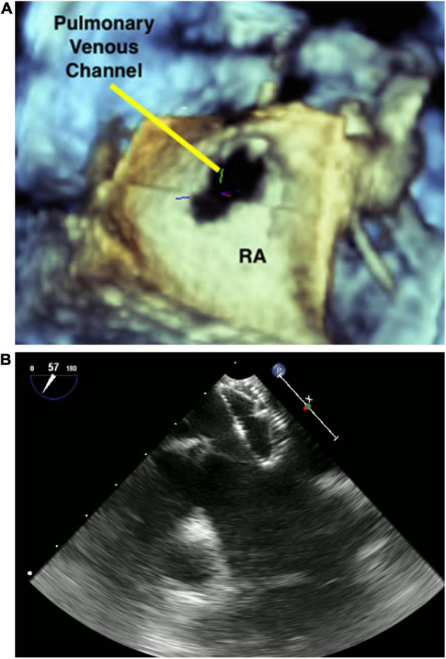
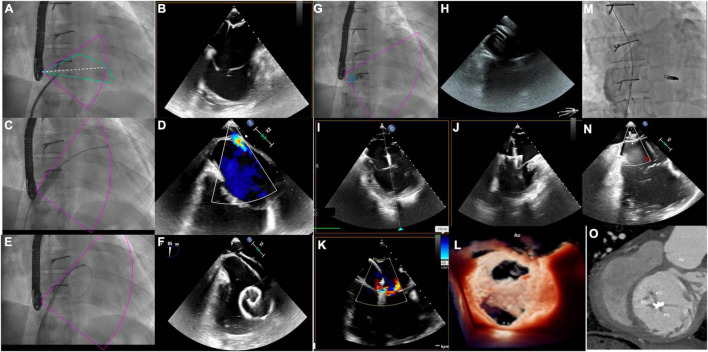
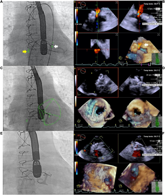
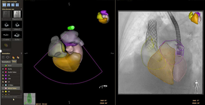
Percutaneous interventions have completely refashioned the management of children with congenital heart diseases (CHD) and the use of non-invasive imaging has become the gold standard to plan and guide these procedures in the modern era. We are now facing a dual challenge to improve the standard of care in low-risk patients, and to shift our strategies from the classic open chest surgery to imaging-guided percutaneous interventions in high-risk patients. Such rapid evolution of ultrasound technologies over the last 20 years have permitted the integration of transthoracic, transesophageal and intracardiac echocardiography into the interventional workflow to improve image guidance and reduce radiation burden from fluoroscopy and angiography. Specifically, miniaturization of transesophageal probe and advances in three-dimensional (3D) imaging techniques have enabled real-time 3D image guidance during complex interventional procedure, In addition, multimodality and fusion imaging techniques harness the strengths of different modalities to enhance understanding of anatomical and spatial relationship between different structures, improving communication and coordination between interventionalists and imaging specialists. In this review, we aim to provide an overview of 3D imaging modalities and multimodal fusion in procedural planning and live guidance of percutaneous interventions. At the present times, 3D imaging can no longer be considered a luxury but a routine clinical tool to improve procedural success and patient outcomes.
经皮介入治疗彻底改变了先天性心脏病(CHD)患儿的治疗方式,在现代,使用非侵入性成像已成为规划和指导这些手术的金标准。我们目前面临着双重挑战,一是提高低风险患者的护理标准,二是将我们的策略从传统的开胸手术转向高风险患者的成像引导经皮介入治疗。在过去20年中,超声技术的迅速发展使得经胸、经食管和心内超声心动图能够整合到介入工作流程中,以改善图像引导并减少荧光透视和血管造影带来的辐射负担。具体而言,经食管探头的小型化和三维(3D)成像技术的进步使得在复杂介入手术过程中能够进行实时3D图像引导。此外,多模态和融合成像技术利用不同模态的优势,增强对不同结构之间解剖和空间关系的理解,改善介入医生和影像专家之间的沟通与协作。在这篇综述中,我们旨在概述3D成像模态以及多模态融合在经皮介入治疗的手术规划和实时引导中的应用。目前,3D成像不再被视为一种奢侈品,而是一种提高手术成功率和患者预后的常规临床工具。