Department of Urology, Kurume University School of Medicine, Kurume, 830-0011, Japan.
Division Microscopic and Development Anatomy, Department of Anatomy, Kurume University School of Medicine, Kurume, 830-0011, Japan.
Sci Rep. 2022 Jun 8;12(1):9484. doi: 10.1038/s41598-022-13245-7.
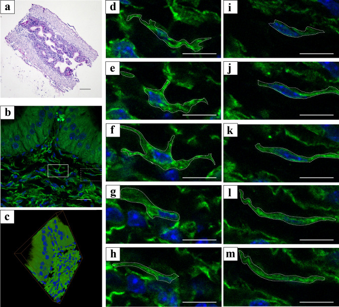
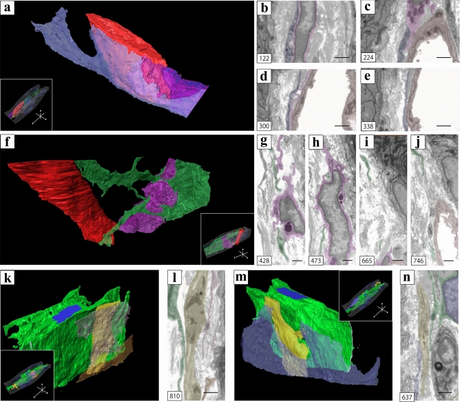
The present study aimed to explore the three-dimensional (3D) ultrastructure of interstitial cells (ICs) within the lamina propria of the murine vas deferens and the spatial relationships between epithelial cells and surrounding cells. Focused ion beam scanning electron microscopy and confocal laser scanning microscopy were performed. ICs within the lamina propria had a flat, sheet-like structure of cytoplasm with multiple cellular processes. In addition, two types of 3D structures that comprised cell processes of flat, sheet-like ICs were observed: one was an accordion fold-like structure and the other was a rod-shaped structure. ICs were located parallel to the epithelium and were connected to each other via gap junctions or adherens junctions. Moreover, multiple sphere-shaped extracellular vesicle-like structures were frequently observed around the ICs. The ICs formed a complex 3D network comprising sheet-like cytoplasm and multiple cell processes with different 3D structures. From this morphological study, we noted that ICs within the lamina propria of murine vas deferens may be involved in signal transmission between the epithelium and smooth muscle cells by physical interaction and by exchanging extracellular vesicles.
本研究旨在探索小鼠输精管固有层内间质细胞(ICs)的三维(3D)超微结构以及上皮细胞与周围细胞之间的空间关系。采用聚焦离子束扫描电子显微镜和共聚焦激光扫描显微镜进行观察。固有层内的 ICs 具有细胞质的扁平片状结构,具有多个细胞突起。此外,观察到两种由扁平片状 ICs 的细胞突起组成的 3D 结构:一种是手风琴折叠样结构,另一种是杆状结构。ICs 平行于上皮排列,通过缝隙连接或黏着连接相互连接。此外,在 ICs 周围经常观察到多个球形细胞外囊泡样结构。ICs 形成了一个复杂的 3D 网络,包括具有不同 3D 结构的扁平片状细胞质和多个细胞突起。从形态学研究中,我们注意到小鼠输精管固有层内的 ICs 可能通过物理相互作用和交换细胞外囊泡参与上皮细胞和平滑肌细胞之间的信号传递。