Department of Neuroscience, UT Southwestern Medical Center, Dallas, TX, 75390, USA.
Lyda Hill Department of Bioinformatics, UT Southwestern Medical Center, Dallas, TX, 75390, USA.
Nat Commun. 2022 Jun 9;13(1):3328. doi: 10.1038/s41467-022-31053-5.
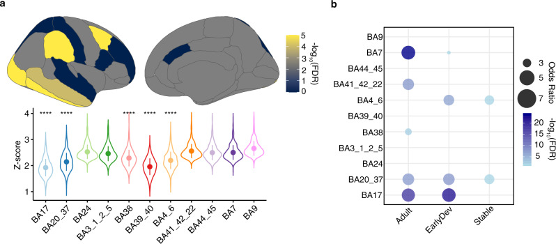
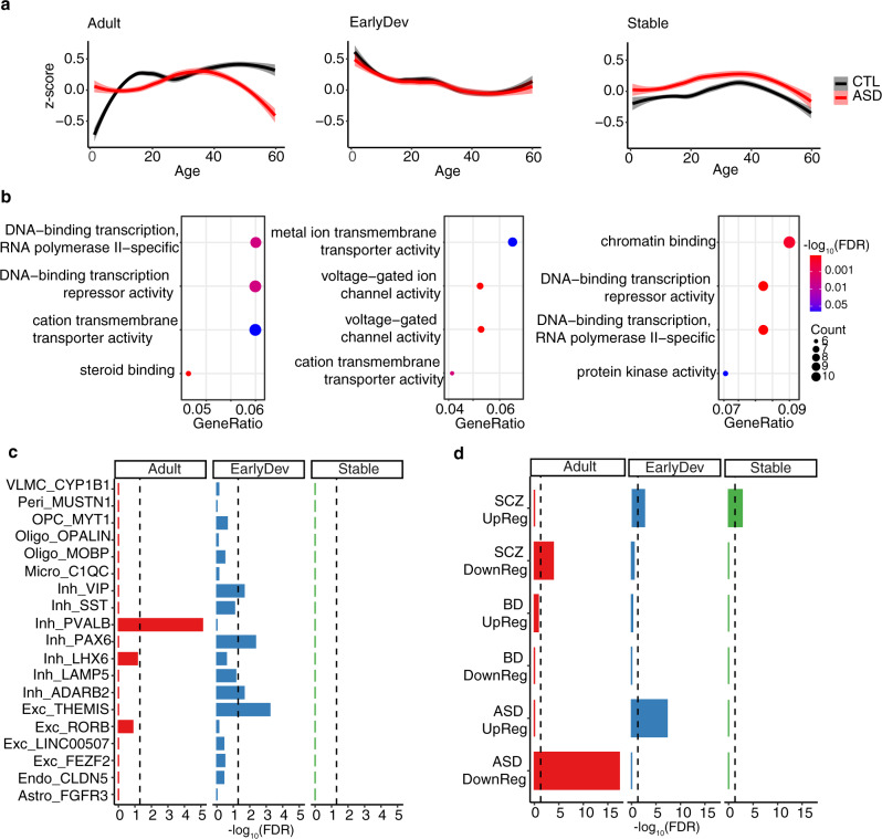
Gene expression covaries with brain activity as measured by resting state functional magnetic resonance imaging (MRI). However, it is unclear how genomic differences driven by disease state can affect this relationship. Here, we integrate from the ABIDE I and II imaging cohorts with datasets of gene expression in brains of neurotypical individuals and individuals with autism spectrum disorder (ASD) with regionally matched brain activity measurements from fMRI datasets. We identify genes linked with brain activity whose association is disrupted in ASD. We identified a subset of genes that showed a differential developmental trajectory in individuals with ASD compared with controls. These genes are enriched in voltage-gated ion channels and inhibitory neurons, pointing to excitation-inhibition imbalance in ASD. We further assessed differences at the regional level showing that the primary visual cortex is the most affected region in ASD. Our results link disrupted brain expression patterns of individuals with ASD to brain activity and show developmental, cell type, and regional enrichment of activity linked genes.
基因表达与静息态功能磁共振成像 (MRI) 测量的大脑活动相关。然而,尚不清楚疾病状态下的基因组差异如何影响这种关系。在这里,我们整合了来自 ABIDE I 和 II 成像队列的数据,以及神经典型个体和自闭症谱系障碍 (ASD) 个体大脑中的基因表达数据集,以及来自 fMRI 数据集的区域匹配大脑活动测量值。我们确定了与大脑活动相关的基因,这些基因在 ASD 中的关联被打破。我们确定了一组在 ASD 个体中与对照组相比表现出不同发育轨迹的基因。这些基因在电压门控离子通道和抑制性神经元中富集,表明 ASD 中存在兴奋抑制失衡。我们进一步评估了区域水平的差异,结果表明初级视觉皮层是 ASD 中受影响最严重的区域。我们的研究结果将 ASD 个体大脑表达模式的紊乱与大脑活动联系起来,并显示出与活动相关基因的发育、细胞类型和区域富集。