• 文献检索
  • 文档翻译
  • 深度研究
  • 学术资讯
  • Suppr Zotero 插件Zotero 插件
  • 邀请有礼
  • 套餐&价格
  • 历史记录
应用&插件
Suppr Zotero 插件Zotero 插件浏览器插件Mac 客户端Windows 客户端微信小程序
定价
高级版会员购买积分包购买API积分包
服务
文献检索文档翻译深度研究API 文档MCP 服务
关于我们
关于 Suppr公司介绍联系我们用户协议隐私条款
关注我们

Suppr 超能文献

核心技术专利:CN118964589B侵权必究
粤ICP备2023148730 号-1Suppr @ 2026

文献检索

告别复杂PubMed语法,用中文像聊天一样搜索,搜遍4000万医学文献。AI智能推荐,让科研检索更轻松。

立即免费搜索

文件翻译

保留排版,准确专业,支持PDF/Word/PPT等文件格式,支持 12+语言互译。

免费翻译文档

深度研究

AI帮你快速写综述,25分钟生成高质量综述,智能提取关键信息,辅助科研写作。

立即免费体验

腹部和骨盆淋巴瘤的并发症:临床和影像学综述。

Complications of lymphoma in the abdomen and pelvis: clinical and imaging review.

机构信息

Department of Diagnostic Radiology, American University of Beirut Medical Center, Riad El-Solh, PO Box 11-0236, Beirut, 1107 2020, Lebanon.

Department of Radiology, Memorial Sloan Kettering Cancer Center, New York, NY, USA.

出版信息

Abdom Radiol (NY). 2022 Aug;47(8):2937-2955. doi: 10.1007/s00261-022-03567-5. Epub 2022 Jun 12.

DOI:10.1007/s00261-022-03567-5
PMID:35690955
原文链接:https://pmc.ncbi.nlm.nih.gov/articles/PMC10509750/
Abstract

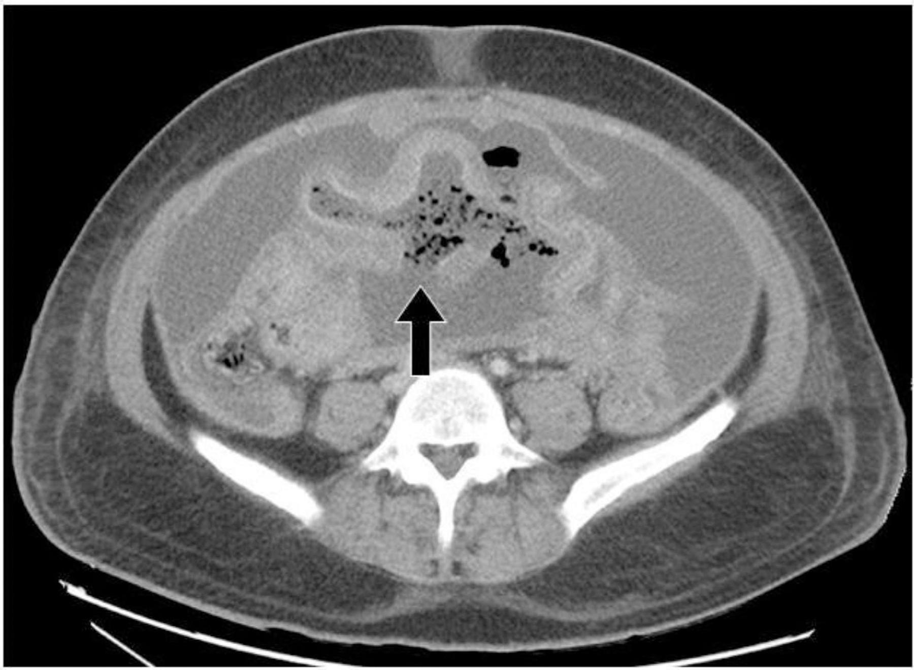
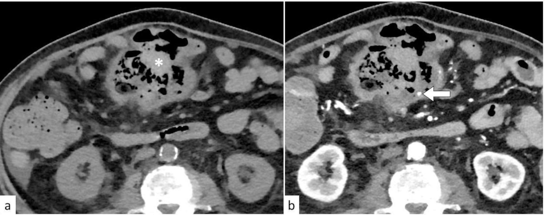
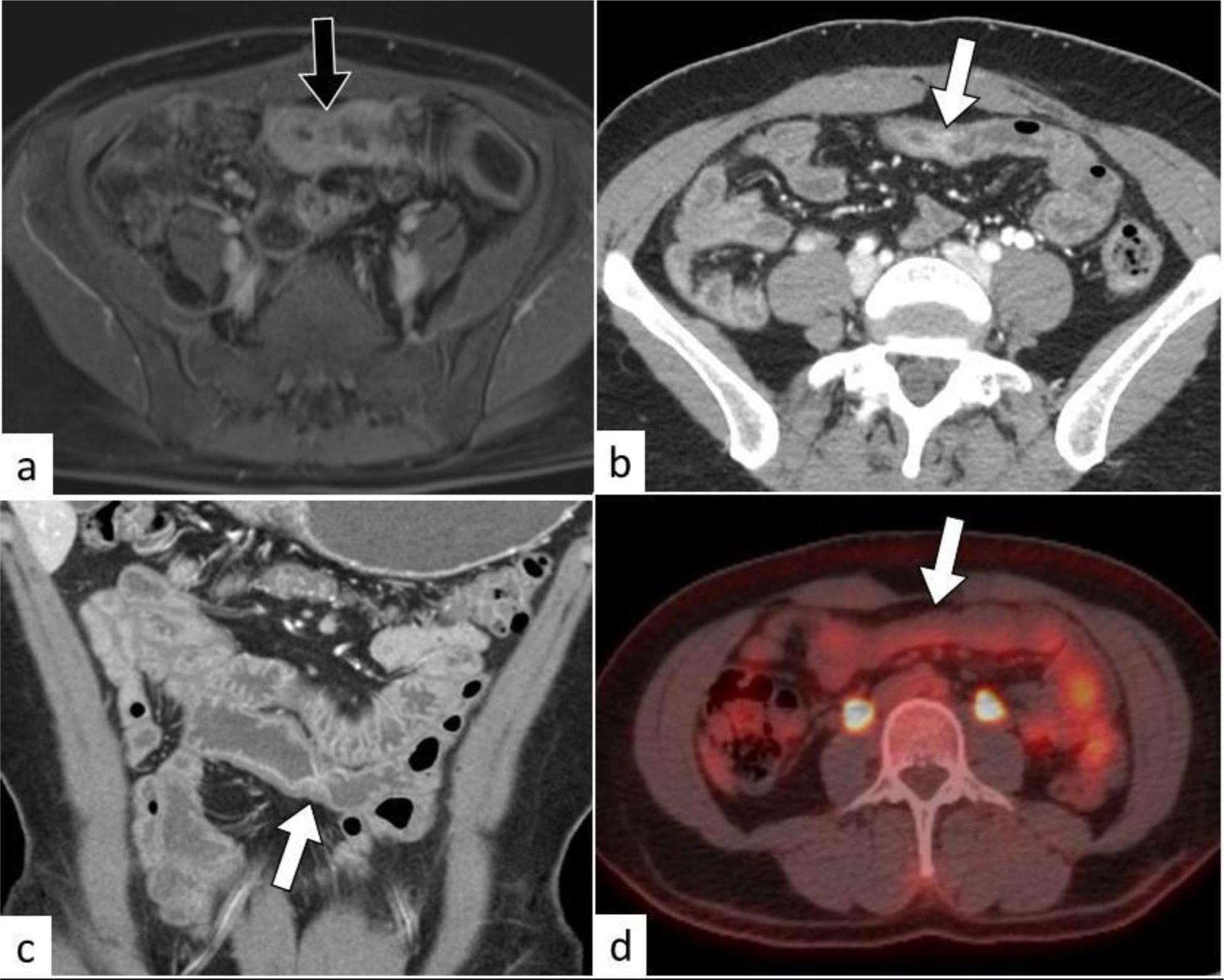
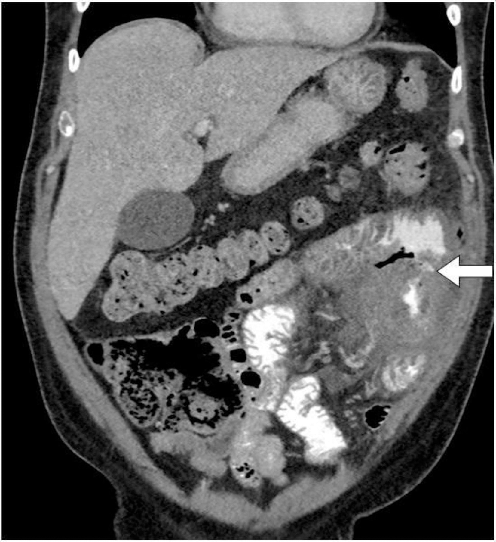
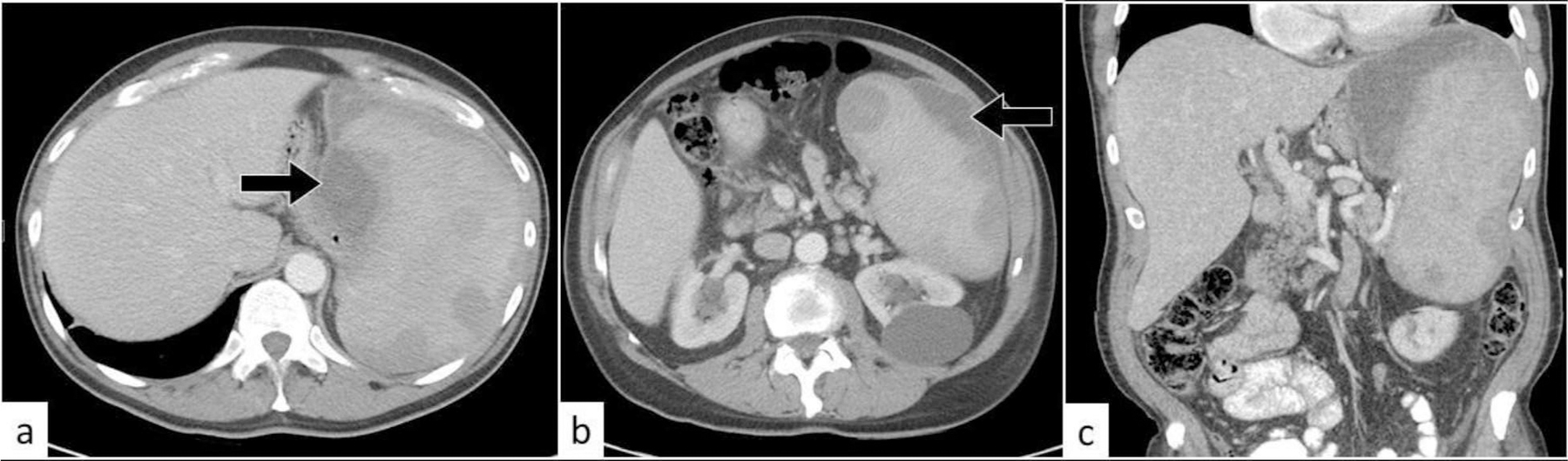
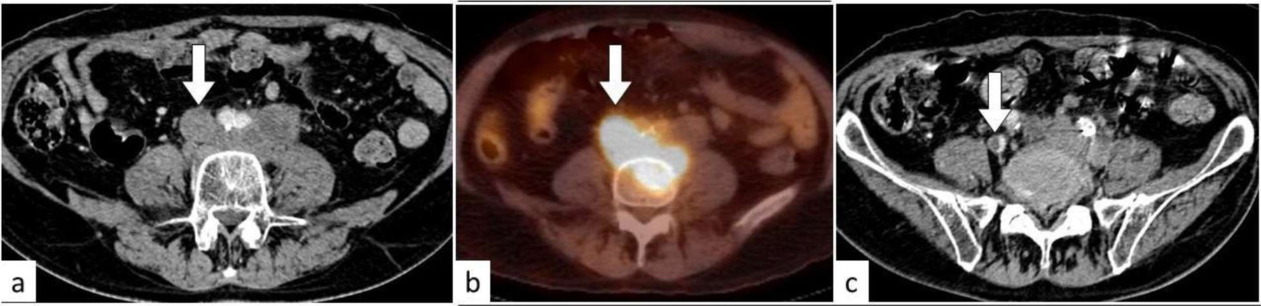
Involvement of the abdomen and pelvis is common in lymphoma. Nodal and extranodal abdominal and pelvic lymphoma may present with various complications. Complications are most common in high-grade lymphomas, especially diffuse large B-cell lymphoma. Complications may occur as the initial manifestation of lymphoma, during treatment course, or late following complete disease remission. Most complications are associated with worse prognosis and increased mortality. Imaging is essential in evaluation of disease extent and diagnosis of complications. Therefore, radiologists should be familiar with the clinical context and imaging features of abdominal and pelvic lymphoma complications. We provide a comprehensive, organ system-based approach, and clinical and imaging review of complications of abdominal and pelvic lymphoma along with radiologic images of illustrated cases of the most commonly encountered complications.

摘要

腹部和骨盆受累在淋巴瘤中很常见。腹部和骨盆的结内和结外淋巴瘤可能会出现各种并发症。并发症在高级别淋巴瘤中最为常见,尤其是弥漫性大 B 细胞淋巴瘤。并发症可能作为淋巴瘤的首发表现,也可能在治疗过程中或完全缓解后晚期出现。大多数并发症与预后较差和死亡率增加有关。影像学检查对于评估疾病范围和诊断并发症至关重要。因此,放射科医生应该熟悉腹部和骨盆淋巴瘤并发症的临床背景和影像学特征。我们提供了一种基于系统的全面方法,对腹部和骨盆淋巴瘤并发症的临床和影像学进行了回顾,并提供了最常见并发症的影像学图片示例。

https://cdn.ncbi.nlm.nih.gov/pmc/blobs/1bbc/10509750/de37e6daf6fb/nihms-1925263-f0029.jpg
https://cdn.ncbi.nlm.nih.gov/pmc/blobs/1bbc/10509750/8149f7371812/nihms-1925263-f0001.jpg
https://cdn.ncbi.nlm.nih.gov/pmc/blobs/1bbc/10509750/f16d8dde7973/nihms-1925263-f0002.jpg
https://cdn.ncbi.nlm.nih.gov/pmc/blobs/1bbc/10509750/419fe9319e6a/nihms-1925263-f0003.jpg
https://cdn.ncbi.nlm.nih.gov/pmc/blobs/1bbc/10509750/55af19da609c/nihms-1925263-f0004.jpg
https://cdn.ncbi.nlm.nih.gov/pmc/blobs/1bbc/10509750/ef7d16580665/nihms-1925263-f0005.jpg
https://cdn.ncbi.nlm.nih.gov/pmc/blobs/1bbc/10509750/68d0b72e706c/nihms-1925263-f0006.jpg
https://cdn.ncbi.nlm.nih.gov/pmc/blobs/1bbc/10509750/a713d362bb7f/nihms-1925263-f0007.jpg
https://cdn.ncbi.nlm.nih.gov/pmc/blobs/1bbc/10509750/dc318fd045db/nihms-1925263-f0008.jpg
https://cdn.ncbi.nlm.nih.gov/pmc/blobs/1bbc/10509750/51b6cf910084/nihms-1925263-f0009.jpg
https://cdn.ncbi.nlm.nih.gov/pmc/blobs/1bbc/10509750/a1e3f9f2fda8/nihms-1925263-f0010.jpg
https://cdn.ncbi.nlm.nih.gov/pmc/blobs/1bbc/10509750/e98736f86ede/nihms-1925263-f0011.jpg
https://cdn.ncbi.nlm.nih.gov/pmc/blobs/1bbc/10509750/bbd6907c94ee/nihms-1925263-f0012.jpg
https://cdn.ncbi.nlm.nih.gov/pmc/blobs/1bbc/10509750/a7f0c129bafe/nihms-1925263-f0013.jpg
https://cdn.ncbi.nlm.nih.gov/pmc/blobs/1bbc/10509750/285e0522d32d/nihms-1925263-f0014.jpg
https://cdn.ncbi.nlm.nih.gov/pmc/blobs/1bbc/10509750/82fb606817f5/nihms-1925263-f0015.jpg
https://cdn.ncbi.nlm.nih.gov/pmc/blobs/1bbc/10509750/3b62829dc4fe/nihms-1925263-f0016.jpg
https://cdn.ncbi.nlm.nih.gov/pmc/blobs/1bbc/10509750/74fd6484338f/nihms-1925263-f0017.jpg
https://cdn.ncbi.nlm.nih.gov/pmc/blobs/1bbc/10509750/a8b5aca73534/nihms-1925263-f0018.jpg
https://cdn.ncbi.nlm.nih.gov/pmc/blobs/1bbc/10509750/1bfa48b39db4/nihms-1925263-f0019.jpg
https://cdn.ncbi.nlm.nih.gov/pmc/blobs/1bbc/10509750/5a52795f72a8/nihms-1925263-f0020.jpg
https://cdn.ncbi.nlm.nih.gov/pmc/blobs/1bbc/10509750/b338ffb9e215/nihms-1925263-f0021.jpg
https://cdn.ncbi.nlm.nih.gov/pmc/blobs/1bbc/10509750/964259404c0b/nihms-1925263-f0022.jpg
https://cdn.ncbi.nlm.nih.gov/pmc/blobs/1bbc/10509750/85b14b580a7a/nihms-1925263-f0023.jpg
https://cdn.ncbi.nlm.nih.gov/pmc/blobs/1bbc/10509750/1d5bd776f7be/nihms-1925263-f0024.jpg
https://cdn.ncbi.nlm.nih.gov/pmc/blobs/1bbc/10509750/747709e30ff0/nihms-1925263-f0025.jpg
https://cdn.ncbi.nlm.nih.gov/pmc/blobs/1bbc/10509750/eb20097093fa/nihms-1925263-f0026.jpg
https://cdn.ncbi.nlm.nih.gov/pmc/blobs/1bbc/10509750/d6e3198145f1/nihms-1925263-f0027.jpg
https://cdn.ncbi.nlm.nih.gov/pmc/blobs/1bbc/10509750/47f2df6c28f8/nihms-1925263-f0028.jpg
https://cdn.ncbi.nlm.nih.gov/pmc/blobs/1bbc/10509750/de37e6daf6fb/nihms-1925263-f0029.jpg

相似文献

1
Complications of lymphoma in the abdomen and pelvis: clinical and imaging review.腹部和骨盆淋巴瘤的并发症:临床和影像学综述。
Abdom Radiol (NY). 2022 Aug;47(8):2937-2955. doi: 10.1007/s00261-022-03567-5. Epub 2022 Jun 12.
2
Cross-sectional imaging of extranodal involvement in abdominopelvic lymphoproliferative malignancies.腹盆腔淋巴增殖性恶性肿瘤结外受累的横断面成像
Radiographics. 2007 Nov-Dec;27(6):1613-34. doi: 10.1148/rg.276065170.
3
Extranodal lymphomas of abdomen and pelvis: imaging findings and differential diagnosis.腹部和骨盆外的淋巴瘤:影像学表现与鉴别诊断。
Abdom Radiol (NY). 2017 Apr;42(4):1096-1112. doi: 10.1007/s00261-016-0964-8.
4
Prognostic impact of abdominal lymph node involvement in diffuse large B-cell lymphoma.弥漫性大 B 细胞淋巴瘤中腹部淋巴结受累的预后影响。
Eur J Haematol. 2020 Mar;104(3):207-213. doi: 10.1111/ejh.13361. Epub 2019 Dec 20.
5
Two Cases of Pelvic Diffuse Large B-Cell Lymphoma.两例盆腔弥漫性大 B 细胞淋巴瘤。
J Investig Med High Impact Case Rep. 2022 Jan-Dec;10:23247096221089505. doi: 10.1177/23247096221089505.
6
Soft-Tissue Sarcomas of the Abdomen and Pelvis: Radiologic-Pathologic Features, Part 2-Uncommon Sarcomas.腹部和盆腔软组织肉瘤:放射学-病理学特征,第2部分-罕见肉瘤
Radiographics. 2017 May-Jun;37(3):797-812. doi: 10.1148/rg.2017160201.
7
Abdominal manifestations of lymphoma: spectrum of imaging features.淋巴瘤的腹部表现:影像特征谱
ISRN Radiol. 2013 Sep 2;2013:483069. doi: 10.5402/2013/483069. eCollection 2013.
8
PET/MRI for the body imager: abdominal and pelvic oncologic applications.用于全身成像的PET/MRI:腹部和盆腔肿瘤学应用
Abdom Imaging. 2015 Aug;40(6):1387-404. doi: 10.1007/s00261-015-0390-3.
9
[Clinical characteristics of 46 pediatric diffuse large B-cell lymphoma and treatment outcome].46例儿童弥漫性大B细胞淋巴瘤的临床特征及治疗结果
Zhonghua Er Ke Za Zhi. 2019 Oct 2;57(10):774-779. doi: 10.3760/cma.j.issn.0578-1310.2019.10.009.
10
The role of MR-PET in abdominal and pelvic oncology.磁共振成像-正电子发射断层显像(MR-PET)在腹部和盆腔肿瘤学中的作用。
Abdom Imaging. 2015 Aug;40(6):1351. doi: 10.1007/s00261-015-0511-z.

引用本文的文献

1
A rare presentation of peritoneal mesothelioma: Esophageal encasement and Sister Mary Joseph nodule.腹膜间皮瘤的罕见表现:食管包绕及玛丽·约瑟夫修女结节。
Radiol Case Rep. 2025 Aug 7;20(11):5407-5410. doi: 10.1016/j.radcr.2025.07.033. eCollection 2025 Nov.
2
Diagnosing Median Arcuate Ligament Syndrome in a Patient With a History of Duodenal Lymphoma: A Case Report.一名有十二指肠淋巴瘤病史患者的正中弓状韧带综合征诊断:病例报告
Cureus. 2025 Feb 27;17(2):e79735. doi: 10.7759/cureus.79735. eCollection 2025 Feb.
3
Recent Advances in Nanobiotechnology for the Treatment of Non-Hodgkin's Lymphoma.

本文引用的文献

1
Imaging findings of spontaneous intraabdominal hemorrhage: neoplastic and non-neoplastic causes.自发性腹腔内出血的影像学表现:肿瘤性和非肿瘤性病因。
Abdom Radiol (NY). 2022 Apr;47(4):1473-1502. doi: 10.1007/s00261-022-03462-z. Epub 2022 Mar 1.
2
Gastrointestinal Bleeding at CT Angiography and CT Enterography: Imaging Atlas and Glossary of Terms.CT 血管造影和 CT 肠造影中的胃肠道出血:成像图谱和术语词汇表。
Radiographics. 2021 Oct;41(6):1632-1656. doi: 10.1148/rg.2021210043.
3
Influence of Severe Gastrointestinal Complications in Primary Gastrointestinal Diffuse Large B-Cell Lymphoma.
纳米生物技术在治疗非霍奇金淋巴瘤中的最新进展。
Mini Rev Med Chem. 2024;24(9):895-907. doi: 10.2174/1389557523666230915103121.
严重胃肠道并发症对原发性胃肠道弥漫性大B细胞淋巴瘤的影响。
Cancer Manag Res. 2021 Feb 4;13:1041-1052. doi: 10.2147/CMAR.S295671. eCollection 2021.
4
Gastric Diffuse Large B-Cell Lymphoma: A Single-Center 9-Year Experience.胃弥漫性大B细胞淋巴瘤:单中心9年经验
Indian J Hematol Blood Transfus. 2021 Jul;37(3):492-496. doi: 10.1007/s12288-020-01391-9. Epub 2021 Jan 2.
5
Clinical spectrum of primary adrenal lymphoma: results of a multicenter cohort study.原发性肾上腺淋巴瘤的临床特征:一项多中心队列研究的结果。
Eur J Endocrinol. 2020 Oct;183(4):453-462. doi: 10.1530/EJE-19-0506.
6
Primary pancreatic lymphoma: Clinical presentation, diagnosis, treatment, and outcome.原发性胰腺淋巴瘤:临床表现、诊断、治疗和预后。
Eur J Haematol. 2020 Oct;105(4):468-475. doi: 10.1111/ejh.13468. Epub 2020 Jul 2.
7
Primary diffuse large B-cell lymphoma presenting as acute appendicitis: A report of 2 cases and a literature review.以急性阑尾炎为表现的原发性弥漫性大B细胞淋巴瘤:2例报告及文献复习
Clin Case Rep. 2020 Jan 7;8(2):293-298. doi: 10.1002/ccr3.2653. eCollection 2020 Feb.
8
Enteral fistula as initial manifestation of primary intestinal lymphoma.肠内瘘作为原发性肠道淋巴瘤的初始表现。
Chin Med J (Engl). 2020 Jan 5;133(1):101-102. doi: 10.1097/CM9.0000000000000598.
9
Imaging of acute abdomen in cancer patients.癌症患者急腹症的影像学表现。
Abdom Radiol (NY). 2020 Aug;45(8):2287-2304. doi: 10.1007/s00261-019-02332-5.
10
Primary renal lymphoma: a population-based study in the United States, 1980-2013.原发性肾淋巴瘤:1980-2013 年美国的一项基于人群的研究。
Sci Rep. 2019 Oct 22;9(1):15125. doi: 10.1038/s41598-019-51635-6.