Nunes-Xavier Caroline E, Mingo Janire, Emaldi Maite, Flem-Karlsen Karine, Mælandsmo Gunhild M, Fodstad Øystein, Llarena Roberto, López José I, Pulido Rafael
Biomarkers in Cancer, Biocruces Bizkaia Health Research Institute, Barakaldo, Spain.
Department of Tumor Biology, Institute for Cancer Research, Oslo University Hospital Radiumhospitalet, Oslo, Norway.
Front Oncol. 2022 May 25;12:873516. doi: 10.3389/fonc.2022.873516. eCollection 2022.
Pyruvate dehydrogenase (PDH) complex converts pyruvate into acetyl-CoA by pyruvate decarboxylation, which drives energy metabolism during cell growth, including prostate cancer (PCa) cell growth. The major catalytic subunit of PDH, PDHA1, is regulated by phosphorylation/dephosphorylation by pyruvate dehydrogenase kinases (PDKs) and pyruvate dehydrogenase phosphatases (PDPs). There are four kinases, PDK1, PDK2, PDK3 and PDK4, which can phosphorylate and inactivate PDH; and two phosphatases, PDP1 and PDP2, that dephosphorylate and activate PDH.
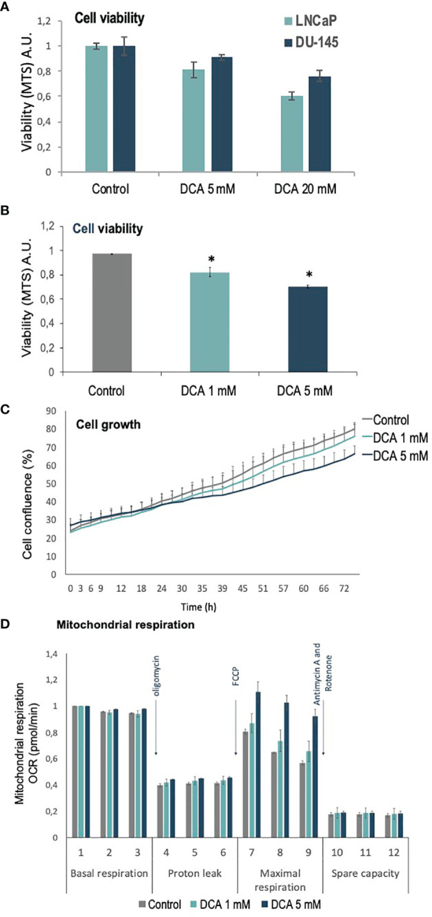
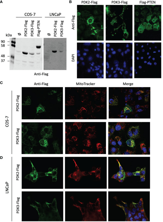
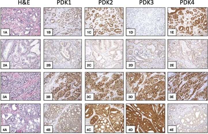
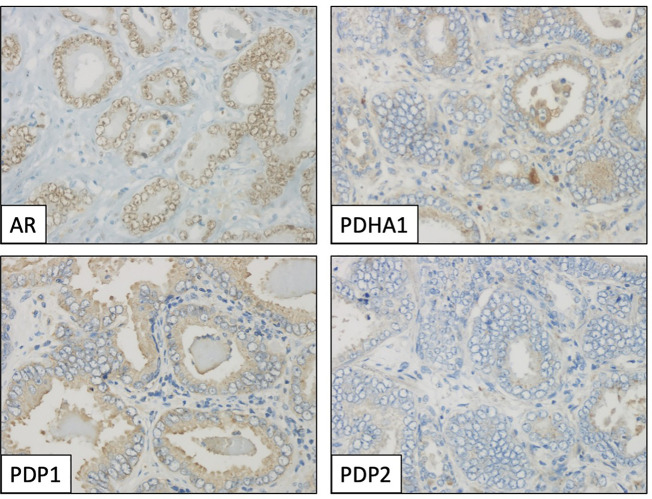
We have analyzed by immunohistochemistry the expression and clinicopathological correlations of PDHA1, PDP1, PDP2, PDK1, PDK2, PDK3, and PDK4, as well as of androgen receptor (AR), in a retrospective PCa cohort of patients. A total of 120 PCa samples of representative tumor areas from all patients were included in tissue microarray (TMA) blocks for analysis. In addition, we studied the subcellular localization of PDK2 and PDK3, and the effects of the PDK inhibitor dichloroacetate (DCA) in the growth, proliferation, and mitochondrial respiration of PCa cells.
We found heterogeneous expression of the PDH complex components in PCa tumors. PDHA1, PDP1, PDK1, PDK2, and PDK4 expression correlated positively with AR expression. A significant correlation of PDK2 immunostaining with biochemical recurrence and disease-free survival was revealed. In PCa tissue specimens, PDK2 displayed cytoplasmic and nuclear immunostaining, whereas PDK1, PDK3 and PDK4 showed mostly cytoplasmic staining. In cells, ectopically expressed PDK2 and PDK3 were mainly localized in mitochondria compartments. An increase in maximal mitochondrial respiration was observed in PCa cells upon PDK inhibition by DCA, in parallel with less proliferative capacity.
Our findings support the notion that expression of specific PDH complex components is related with AR signaling in PCa tumors. Furthermore, PDK2 expression associated with poor PCa prognosis. This highlights a potential for PDH complex components as targets for intervention in PCa.
丙酮酸脱氢酶(PDH)复合物通过丙酮酸脱羧作用将丙酮酸转化为乙酰辅酶A,这在包括前列腺癌(PCa)细胞生长在内的细胞生长过程中驱动能量代谢。PDH的主要催化亚基PDHA1受丙酮酸脱氢酶激酶(PDK)和丙酮酸脱氢酶磷酸酶(PDP)的磷酸化/去磷酸化调节。有四种激酶,即PDK1、PDK2、PDK3和PDK4,它们可使PDH磷酸化并使其失活;还有两种磷酸酶,即PDP1和PDP2,它们使PDH去磷酸化并激活PDH。
我们通过免疫组织化学分析了一组前列腺癌患者回顾性队列中PDHA1、PDP1、PDP2、PDK1、PDK2、PDK3和PDK4以及雄激素受体(AR)的表达及其与临床病理的相关性。将所有患者具有代表性肿瘤区域的总共120个PCa样本纳入组织微阵列(TMA)块进行分析。此外,我们研究了PDK2和PDK3的亚细胞定位,以及PDK抑制剂二氯乙酸(DCA)对PCa细胞生长、增殖和线粒体呼吸的影响。
我们发现PCa肿瘤中PDH复合物成分表达存在异质性。PDHA1、PDP1、PDK1、PDK2和PDK4的表达与AR表达呈正相关。PDK2免疫染色与生化复发和无病生存期之间存在显著相关性。在PCa组织标本中,PDK2显示出细胞质和细胞核免疫染色,而PDK1、PDK3和PDK4大多显示细胞质染色。在细胞中,异位表达的PDK2和PDK3主要定位于线粒体区室。在用DCA抑制PDK后,PCa细胞中的最大线粒体呼吸增加,同时增殖能力降低。
我们的研究结果支持以下观点,即特定PDH复合物成分的表达与PCa肿瘤中的AR信号传导相关。此外,PDK2表达与PCa预后不良相关。这突出了PDH复合物成分作为PCa干预靶点的潜力。