Department of Diagnostic and Interventional Neuroradiology, University Medical Center Hamburg-Eppendorf, Lokstedter Weg 104, 20251, Hamburg, Germany.
Department of Pediatrics, University Medical Center Hamburg-Eppendorf, Hamburg, Germany.
Neuroradiology. 2022 Oct;64(10):2059-2067. doi: 10.1007/s00234-022-02988-9. Epub 2022 Jun 14.
Grey matter (GM) atrophy due to neuronal loss is a striking feature of patients with CLN3 disease. A precise and quantitative description of disease progression is needed in order to establish an evaluation tool for current and future experimental treatments. In order to develop a quantitative marker to measure brain volume outcome, we analysed the longitudinal volumetric development of GM, white matter (WM) and lateral ventricles and correlated those with the clinical course.
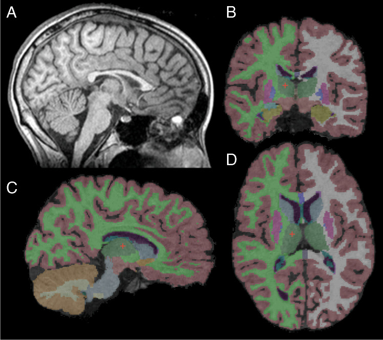
One hundred twenty-two MRI scans of 35 patients (21 females; 14 males; age 15.3 ± 4.8 years) with genetically confirmed CLN3 disease were performed. A three-dimensional T1-weighted sequence was acquired with whole brain coverage. Volumetric segmentation of the brain was performed with the FreeSurfer image analysis suite. The clinical severity was assessed by the Hamburg jNCL score, a disease-specific scoring system.
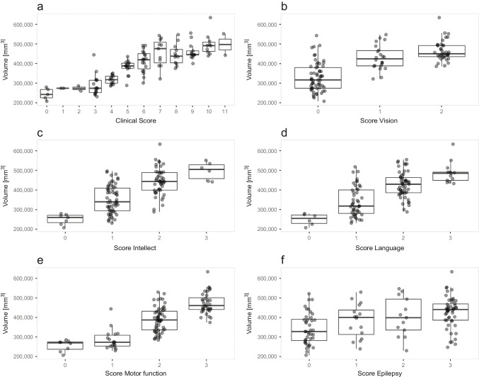
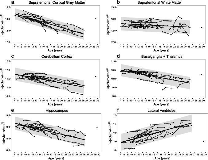
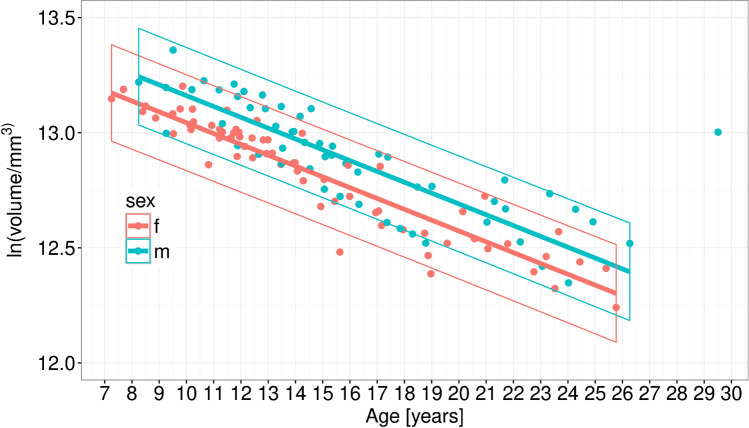
The volumes of supratentorial cortical GM and supratentorial WM, cerebellar GM, basal ganglia/thalamus and hippocampus significantly (r = - 0.86 to - 0.69, p < 0.0001) decreased with age, while the lateral ventricle volume increased (r = 0.68, p < 0.0001). Supratentorial WM volume correlated poorer with age (r = - 0.56, p = 0.0001). Supratentorial cortical GM volume showed the steepest (4.6% (± 0.2%)) and most uniform decrease with strongest correlation with age (r = - 0.86, p < 0.0001). In addition, a strong correlation with disease specific clinical scoring existed for the supratentorial cortical GM volume (r = 0.85, p = < 0.0001).
Supratentorial cortical GM volume is a sensitive parameter for assessment of disease progression even in early and late disease stages and represents a potential reliable outcome measure for evaluation of experimental therapies.
由于神经元丢失导致的灰质(GM)萎缩是 CLN3 病患者的一个显著特征。为了建立当前和未来实验治疗的评估工具,需要对疾病进展进行精确和定量描述。为了开发一种定量标记物来测量脑容量结果,我们分析了 GM、白质(WM)和侧脑室的纵向体积发育,并将其与临床病程相关联。
对 35 名经基因证实的 CLN3 病患者(21 名女性;14 名男性;年龄 15.3 ± 4.8 岁)进行了 122 次 MRI 扫描。使用全脑覆盖的三维 T1 加权序列进行采集。使用 FreeSurfer 图像分析套件对大脑进行容积分割。临床严重程度通过汉堡 jNCL 评分(一种特定于疾病的评分系统)进行评估。
大脑皮质 GM 和大脑皮质 WM、小脑 GM、基底节/丘脑和海马的体积明显(r= −0.86 至−0.69,p<0.0001)随年龄而减少,而侧脑室体积增加(r=0.68,p<0.0001)。大脑皮质 WM 体积与年龄的相关性较差(r=−0.56,p=0.0001)。大脑皮质 GM 体积显示出最陡峭(4.6%(±0.2%))和最均匀的下降,与年龄的相关性最强(r=−0.86,p<0.0001)。此外,大脑皮质 GM 体积与特定于疾病的临床评分之间存在很强的相关性(r=0.85,p= <0.0001)。
大脑皮质 GM 体积是评估疾病进展的敏感参数,即使在早期和晚期疾病阶段也是如此,并且代表了评估实验治疗的潜在可靠结果测量指标。