• 文献检索
  • 文档翻译
  • 深度研究
  • 学术资讯
  • Suppr Zotero 插件Zotero 插件
  • 邀请有礼
  • 套餐&价格
  • 历史记录
应用&插件
Suppr Zotero 插件Zotero 插件浏览器插件Mac 客户端Windows 客户端微信小程序
定价
高级版会员购买积分包购买API积分包
服务
文献检索文档翻译深度研究API 文档MCP 服务
关于我们
关于 Suppr公司介绍联系我们用户协议隐私条款
关注我们

Suppr 超能文献

核心技术专利:CN118964589B侵权必究
粤ICP备2023148730 号-1Suppr @ 2026

文献检索

告别复杂PubMed语法,用中文像聊天一样搜索,搜遍4000万医学文献。AI智能推荐,让科研检索更轻松。

立即免费搜索

文件翻译

保留排版,准确专业,支持PDF/Word/PPT等文件格式,支持 12+语言互译。

免费翻译文档

深度研究

AI帮你快速写综述,25分钟生成高质量综述,智能提取关键信息,辅助科研写作。

立即免费体验

EpiSmart上皮交联术后圆锥角膜和手术后角膜扩张的角膜厚度测量评估

Pachymetric Assessment After EpiSmart Epithelium-on Cross-Linking for Keratoconus and Post-Surgical Ectasia.

作者信息

Vaidya Neel S, Daneshmand Arvin, Epstein Randy J, Majmudar Parag A, Belin Michael W, Parsons Edward C, Rubinfeld Roy S

机构信息

Chicago Cornea Consultants, Highland Park, IL, USA.

Department of Ophthalmology, Rush University Medical Center, Chicago, IL, USA.

出版信息

Clin Ophthalmol. 2022 Jun 8;16:1829-1835. doi: 10.2147/OPTH.S359710. eCollection 2022.

DOI:10.2147/OPTH.S359710
PMID:35702687
原文链接:https://pmc.ncbi.nlm.nih.gov/articles/PMC9188777/
Abstract

PURPOSE

To assess the change in corneal pachymetry after a novel epithelium-on (EpiSmart) corneal crosslinking procedure (CXL).

METHODS

Eyes treated as part of the open-label, non-controlled arm of the study "Collagen Crosslinking with Ultraviolet-A in Asymmetric Corneas" (NCT01097447) were examined at baseline, 3-, 6- and 12-months post-CXL. Thinnest pachymetry readings based on Pentacam (OCULUS GmbH, Wetzlar, Germany) were recorded.

RESULTS

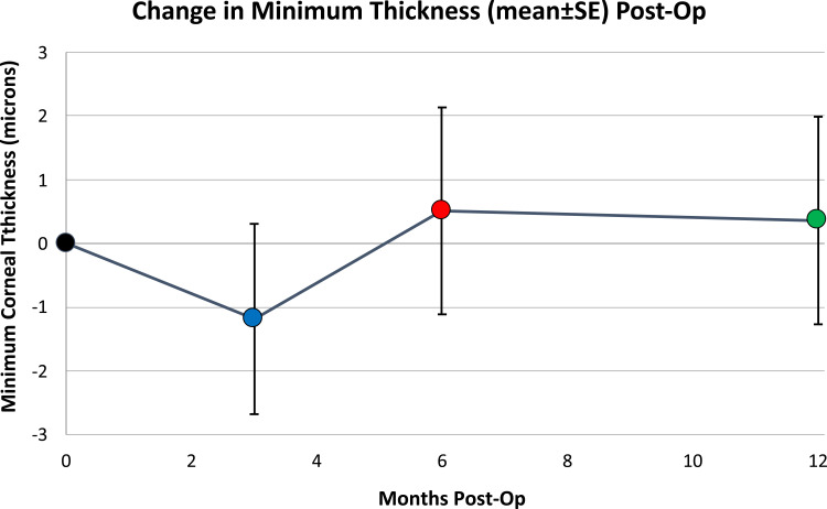
A total of 101 eyes met the study inclusion criteria. Thinnest pachymetric readings at baseline averaged 451 ± 50 microns. The mean (± SD) minimum thickness was 450 ± 46 microns at 3 months, 452 ± 47 microns at 6 months, and 451 ± 48 microns at 12 months post-CXL. The changes from baseline (mean ± SE) at 3, 6, and 12 months post-CXL were -1.2 ± 1.5 microns, 0.5 ± 1.6 microns, and 0.4 ± 1.6 microns, respectively. Student's t-tests showed no statistically significant change in pachymetry from baseline for any exam period.

CONCLUSION

This study demonstrated that, after EpiSmart epithelium-on CXL, there was no substantial corneal thinning observable on Scheimpflug tomography out to 12 months.

摘要

目的

评估一种新型表层角膜交联术(EpiSmart)后角膜厚度测量值的变化。

方法

在“非对称角膜的紫外线A胶原交联术”(NCT01097447)研究的开放标签、非对照组中接受治疗的眼睛,在角膜交联术后基线、3个月、6个月和12个月时进行检查。记录基于Pentacam(德国韦茨拉尔的OCULUS GmbH公司)的最薄角膜厚度测量值。

结果

共有101只眼睛符合研究纳入标准。基线时最薄角膜厚度测量值平均为451±50微米。角膜交联术后3个月时平均(±标准差)最小厚度为450±46微米,6个月时为452±47微米,12个月时为451±48微米。角膜交联术后3个月、6个月和12个月时相对于基线的变化(平均值±标准误)分别为-1.2±1.5微米、0.5±1.6微米和0.4±1.6微米。学生t检验显示,在任何检查时间段,角膜厚度测量值与基线相比均无统计学上的显著变化。

结论

本研究表明,在进行EpiSmart表层角膜交联术后,至12个月时,在Scheimpflug断层扫描中未观察到明显的角膜变薄。

https://cdn.ncbi.nlm.nih.gov/pmc/blobs/316b/9188777/060c38adc755/OPTH-16-1829-g0003.jpg
https://cdn.ncbi.nlm.nih.gov/pmc/blobs/316b/9188777/189353729dab/OPTH-16-1829-g0001.jpg
https://cdn.ncbi.nlm.nih.gov/pmc/blobs/316b/9188777/2c2db0752d92/OPTH-16-1829-g0002.jpg
https://cdn.ncbi.nlm.nih.gov/pmc/blobs/316b/9188777/060c38adc755/OPTH-16-1829-g0003.jpg
https://cdn.ncbi.nlm.nih.gov/pmc/blobs/316b/9188777/189353729dab/OPTH-16-1829-g0001.jpg
https://cdn.ncbi.nlm.nih.gov/pmc/blobs/316b/9188777/2c2db0752d92/OPTH-16-1829-g0002.jpg
https://cdn.ncbi.nlm.nih.gov/pmc/blobs/316b/9188777/060c38adc755/OPTH-16-1829-g0003.jpg

相似文献

1
Pachymetric Assessment After EpiSmart Epithelium-on Cross-Linking for Keratoconus and Post-Surgical Ectasia.EpiSmart上皮交联术后圆锥角膜和手术后角膜扩张的角膜厚度测量评估
Clin Ophthalmol. 2022 Jun 8;16:1829-1835. doi: 10.2147/OPTH.S359710. eCollection 2022.
2
Transepithelial versus epithelium-off corneal crosslinking for progressive keratoconus.经上皮角膜交联与角膜上皮瓣下角膜交联治疗进行性圆锥角膜的比较。
Cochrane Database Syst Rev. 2021 Mar 23;3(3):CD013512. doi: 10.1002/14651858.CD013512.pub2.
3
Corneal thickness measurements using time-domain anterior segment OCT, ultrasound, and Scheimpflug tomographer pachymetry before and after corneal cross-linking for keratoconus.角膜交联术治疗圆锥角膜前后应用时域眼前节 OCT、超声和 Scheimpflug 角膜地形图仪测量角膜厚度。
J Refract Surg. 2012 Aug;28(8):562-6. doi: 10.3928/1081597X-20120703-02. Epub 2012 Jul 13.
4
Epithelium-off corneal collagen cross-linking versus transepithelial cross-linking for pediatric keratoconus.去上皮角膜胶原交联术与经上皮角膜交联术治疗儿童圆锥角膜的比较。
Cornea. 2013 May;32(5):597-601. doi: 10.1097/ICO.0b013e31826cf32d.
5
Corneal densitometry after accelerated corneal collagen cross-linking in progressive keratoconus.进行性圆锥角膜加速角膜胶原交联后的角膜密度测定
Int Ophthalmol. 2019 Apr;39(4):765-775. doi: 10.1007/s10792-018-0876-4. Epub 2018 Mar 26.
6
Assessment of the tomographic values in keratoconic eyes after collagen crosslinking procedure.胶原交联术后圆锥角膜眼断层扫描值的评估
Med Arch. 2015 Apr;69(2):91-4. doi: 10.5455/medarh.2015.69.91-94. Epub 2015 Apr 6.
7
Three-year follow-up in advanced pediatric keratoconus: thin corneas may not have pachymetry properly assessed after crosslinking.儿童晚期圆锥角膜的三年随访:交联术后薄角膜可能未得到准确的角膜厚度测量评估。
Int J Ophthalmol. 2020 Oct 18;13(10):1561-1566. doi: 10.18240/ijo.2020.10.08. eCollection 2020.
8
Long-term visual, refractive, tomographic and aberrometric outcomes of corneal collagen crosslinking (CXL) with or without hypoosmolar riboflavin solution in the treatment of progressive keratoconus patients with thin corneas.角膜胶原交联术(CXL)联合或不联合低渗核黄素溶液治疗薄角膜进展性圆锥角膜患者的长期视力、屈光、断层和像差结果。
Graefes Arch Clin Exp Ophthalmol. 2022 Apr;260(4):1225-1235. doi: 10.1007/s00417-021-05314-w. Epub 2021 Nov 27.
9
Pachymetry and Elevation Back Map Changes in Keratoconus Patients After Crosslinking Procedure.交联术后圆锥角膜患者角膜厚度和高度背侧地图变化。
Med Arch. 2020 Apr;74(2):105-108. doi: 10.5455/medarh.2020.74.105-108.
10
Morphological and functional correlations in riboflavin UV A corneal collagen cross-linking for keratoconus.核黄素 UVA 角膜胶原交联术治疗圆锥角膜的形态与功能相关性。
Acta Ophthalmol. 2012 May;90(3):259-65. doi: 10.1111/j.1755-3768.2010.01890.x. Epub 2010 Apr 23.

引用本文的文献

1
Recent advances in medicinal compounds related to corneal crosslinking.与角膜交联相关的药用化合物的最新进展。
Front Pharmacol. 2023 Sep 29;14:1232591. doi: 10.3389/fphar.2023.1232591. eCollection 2023.

本文引用的文献

1
Corneal Stability and Visual Acuity 1 Year After Corneal Cross-linking Assessed Using the ABCD Keratoconus Staging System.角膜交联术后 1 年应用 ABCD 圆锥角膜分级系统评估角膜稳定性和视力
J Refract Surg. 2021 Oct;37(10):700-706. doi: 10.3928/1081597X-20210712-09. Epub 2021 Oct 1.
2
Keratoconus: Diagnosis and Staging.圆锥角膜:诊断与分期。
Cornea. 2022 Jan 1;41(1):1-11. doi: 10.1097/ICO.0000000000002781.
3
The Effect of Sodium Iodide on Stromal Loading, Distribution and Degradation of Riboflavin in a Rabbit Model of Transepithelial Corneal Crosslinking.
碘化钠对兔上皮下角膜交联模型中核黄素基质负载、分布及降解的影响
Clin Ophthalmol. 2021 May 11;15:1985-1994. doi: 10.2147/OPTH.S300886. eCollection 2021.
4
Comparative Efficacy and Safety of Standard Versus Accelerated Corneal Crosslinking for Keratoconus: 1-Year Outcomes From the Save Sight Keratoconus Registry Study.圆锥角膜标准与加速角膜交联术的疗效与安全性比较:来自拯救视力圆锥角膜登记研究的1年结果
Cornea. 2021 Dec 1;40(12):1581-1589. doi: 10.1097/ICO.0000000000002747.
5
Selective transepithelial ablation with simultaneous accelerated corneal crosslinking for corneal regularization of keratoconus: STARE-X protocol.选择性经上皮消融联合加速角膜交联术治疗圆锥角膜的角膜规则化:STARE-X 方案。
J Cataract Refract Surg. 2021 Nov 1;47(11):1403-1410. doi: 10.1097/j.jcrs.0000000000000640.
6
Transepithelial versus Epithelium-off Corneal Collagen Cross-linking for Corneal Ectasia: A Systematic Review and Meta-analysis.经上皮与去上皮角膜胶原交联治疗角膜扩张症的比较:系统评价和荟萃分析。
Ophthalmology. 2021 Aug;128(8):1150-1160. doi: 10.1016/j.ophtha.2020.12.023. Epub 2020 Dec 28.
7
Pachymetry and Elevation Back Map Changes in Keratoconus Patients After Crosslinking Procedure.交联术后圆锥角膜患者角膜厚度和高度背侧地图变化。
Med Arch. 2020 Apr;74(2):105-108. doi: 10.5455/medarh.2020.74.105-108.
8
Transepithelial versus epithelium-off corneal crosslinking for corneal ectasia.经上皮与去上皮角膜交联术治疗角膜扩张症。
J Cataract Refract Surg. 2018 Dec;44(12):1507-1516. doi: 10.1016/j.jcrs.2018.08.021. Epub 2018 Oct 9.
9
Corneal crosslinking without epithelial removal.角膜交联术无需去除上皮。
J Cataract Refract Surg. 2018 Nov;44(11):1363-1370. doi: 10.1016/j.jcrs.2018.07.029. Epub 2018 Sep 15.
10
Quantitative analysis of corneal stromal riboflavin concentration without epithelial removal.定量分析角膜基质内的核黄素浓度,无需去除上皮细胞。
J Cataract Refract Surg. 2018 Feb;44(2):237-242. doi: 10.1016/j.jcrs.2018.01.010. Epub 2018 Mar 8.