The Shapiro Family Laboratory of Viral Oncology and Aging Research, UCLA School of Dentistry, Los Angeles, CA 90095, USA.
Section of Oral Biology, UCLA School of Dentistry, Los Angeles, CA 90095, USA.
Int J Mol Med. 2022 Aug;50(2). doi: 10.3892/ijmm.2022.5159. Epub 2022 Jun 15.
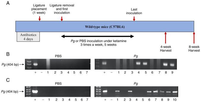
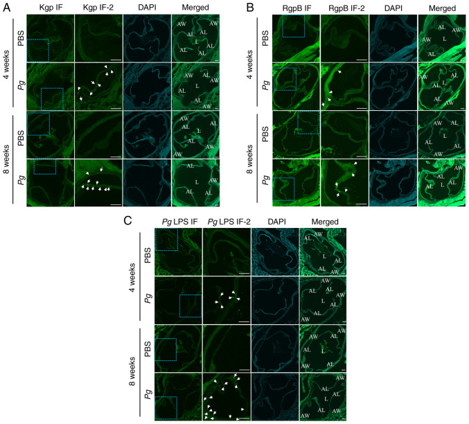
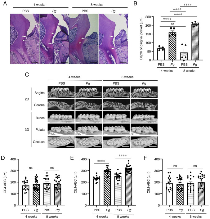
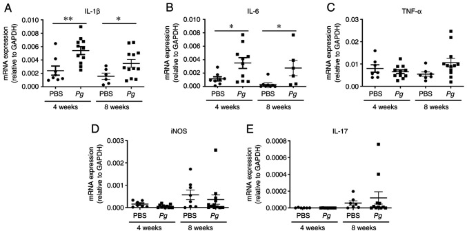
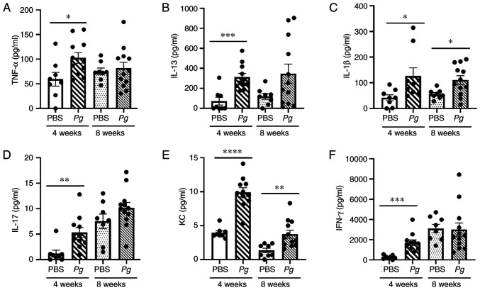
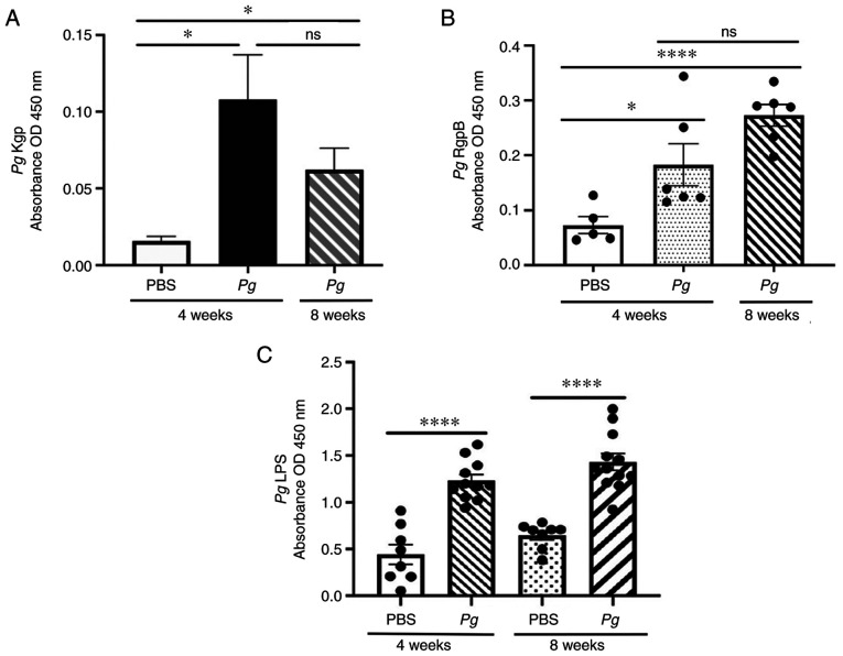
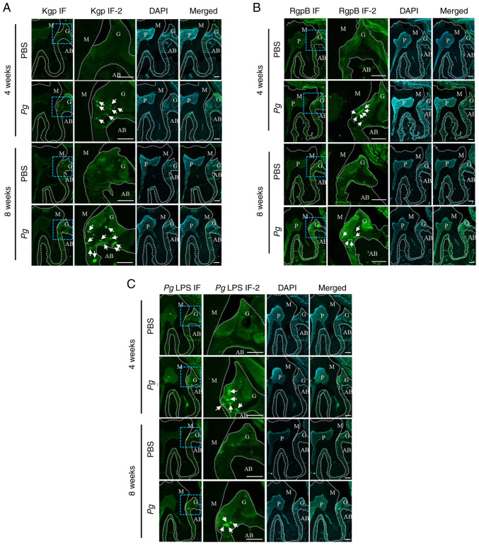
(), one of the 'red‑complex' perio‑pathogens known to play a critical role in the development of periodontitis, has been used in various animal models to mimic human bacteria‑induced periodontitis. In order to achieve a more realistic animal model of human infection, the present study investigated whether repeated small‑volume topical applications of directly into the gingival pocket can induce local infection, including periodontitis and systemic vascular inflammation in wild‑type mice. Freshly cultured was topically applied directly into the gingival pocket of the second molars for 5 weeks (3 times/week). After the final application, the mice were left in cages for 4 or 8 weeks and sacrificed. The status of colony formation in the pocket, gingival inflammation, alveolar bone loss, the expression levels of pro‑inflammatory cytokines in the serum and aorta, the presence of anti‑ lipopolysaccharide (LPS) and gingipain (Kpg and RgpB) antibodies in the serum, as well as the accumulation of LPS and gingipain aggregates in the gingiva and arterial wall were evaluated. The topical application of into the gingival pocket induced the following local and systemic pathohistological changes in mice when examined at 4 or 8 weeks after the final topical application: colonization in the majority of gingival pockets; increased gingival pocket depths; gingival inflammation indicated by the increased expression of TNF‑α, IL‑6 and IL‑1β; significant loss of alveolar bone at the sites of topical application; and increased levels of pro‑inflammatory cytokines, such as TNF‑α, IL‑1β, IL‑17, IL‑13, KC and IFN‑γ in the serum in comparison to those from mice receiving PBS. In addition, the application/colonization model induced anti‑ LPS and gingipain antibodies in serum, as well as the accumulation of LPS and gingipain aggregates in the gingivae and arterial walls. To the best of our knowledge, this mouse model represents the first example of creating a more sustained local infection in the gingival tissues of wild‑type mice and may prove to be useful for the investigation of the more natural and complete pathogenesis of the bacteria in the development of local oral and systemic diseases, such as atherosclerosis. It may also be useful for the determination of a treatment/prevention/efficacy model associated with ‑induced colonization periodontitis in mice.
牙龈卟啉单胞菌是已知的“红色复合体”牙周病原体之一,在牙周炎的发展中起着关键作用,已被用于各种动物模型来模拟人类细菌诱导的牙周炎。为了实现更逼真的人类感染动物模型,本研究探讨了是否可以通过反复将小体积的牙龈卟啉单胞菌直接涂抹到牙龈袋中来诱导局部感染,包括野生型小鼠的牙周炎和全身血管炎症。将新鲜培养的牙龈卟啉单胞菌直接涂抹到第二磨牙的牙龈袋中,每周 3 次,共 5 周。末次给药后,将小鼠置于笼中 4 或 8 周后处死。评估口袋中菌斑形成情况、牙龈炎症、牙槽骨丧失、血清和主动脉中促炎细胞因子的表达水平、血清中抗脂多糖(LPS)和牙龈蛋白酶(Kpg 和 RgpB)抗体的存在情况,以及牙龈和动脉壁中 LPS 和牙龈蛋白酶聚集体的积累情况。当在末次局部牙龈卟啉单胞菌给药后 4 或 8 周检查时,将牙龈卟啉单胞菌直接涂抹到牙龈袋中可诱导小鼠出现以下局部和全身组织病理学变化:大多数牙龈袋中有定植;牙龈袋深度增加;TNF-α、IL-6 和 IL-1β 表达增加表明牙龈炎症;在局部牙龈卟啉单胞菌应用部位牙槽骨显著丧失;与接受 PBS 的小鼠相比,血清中促炎细胞因子(如 TNF-α、IL-1β、IL-17、IL-13、KC 和 IFN-γ)水平升高。此外,牙龈卟啉单胞菌应用/定植模型诱导了血清中抗 LPS 和牙龈蛋白酶抗体,以及牙龈和动脉壁中 LPS 和牙龈蛋白酶聚集体的积累。据我们所知,这种小鼠模型代表了在野生型小鼠牙龈组织中建立更持续局部感染的首例实例,并且可能有助于研究细菌在局部口腔和全身疾病(如动脉粥样硬化)发展中的更自然和完整的发病机制。它也可能有助于确定与牙龈卟啉单胞菌定植牙周炎相关的治疗/预防/疗效模型。