Immune System Development and Function Unit, Centro de Biología Molecular Severo Ochoa, Consejo Superior de Investigaciones Científicas (CSIC), Universidad Autónoma de Madrid (UAM), Madrid, Spain.
Department of Biomedicine and University Children's Hospital of Basel, University of Basel, Basel, Switzerland.
Front Immunol. 2022 May 30;13:867302. doi: 10.3389/fimmu.2022.867302. eCollection 2022.
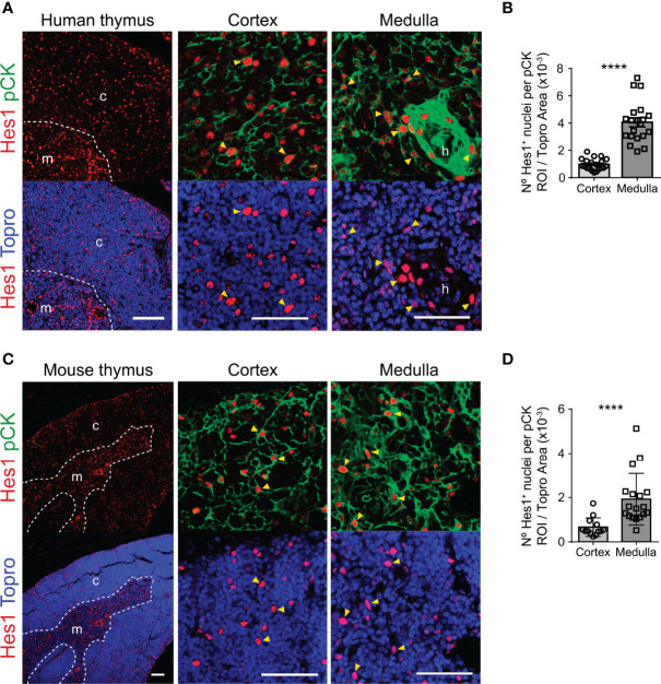
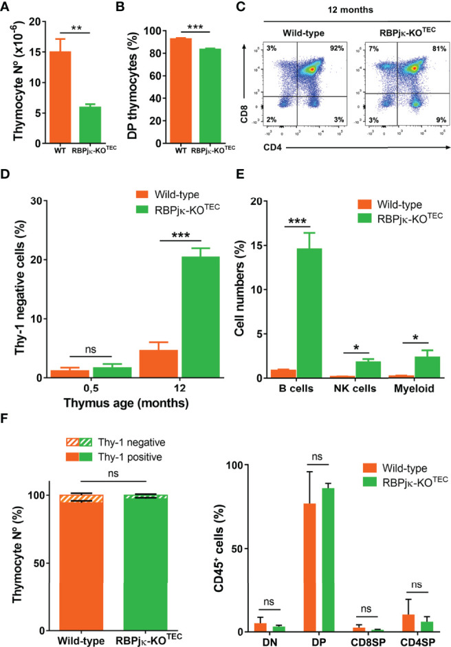
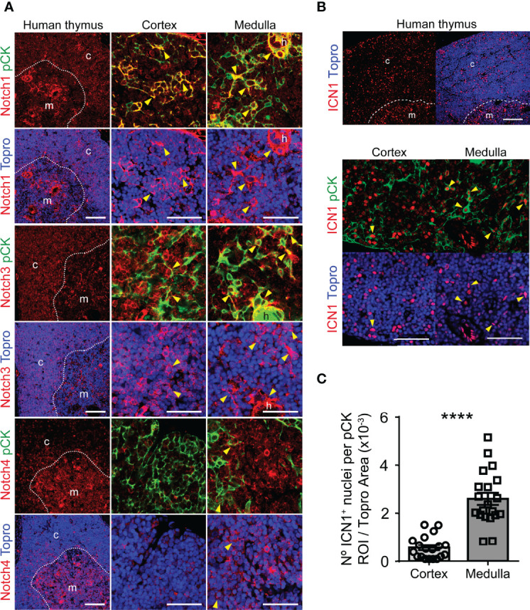
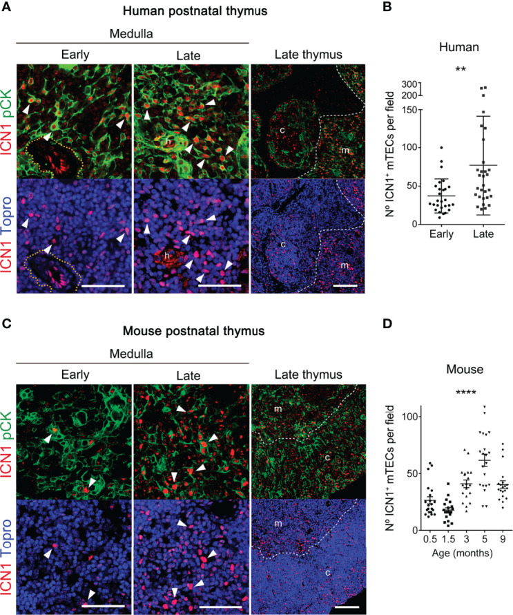
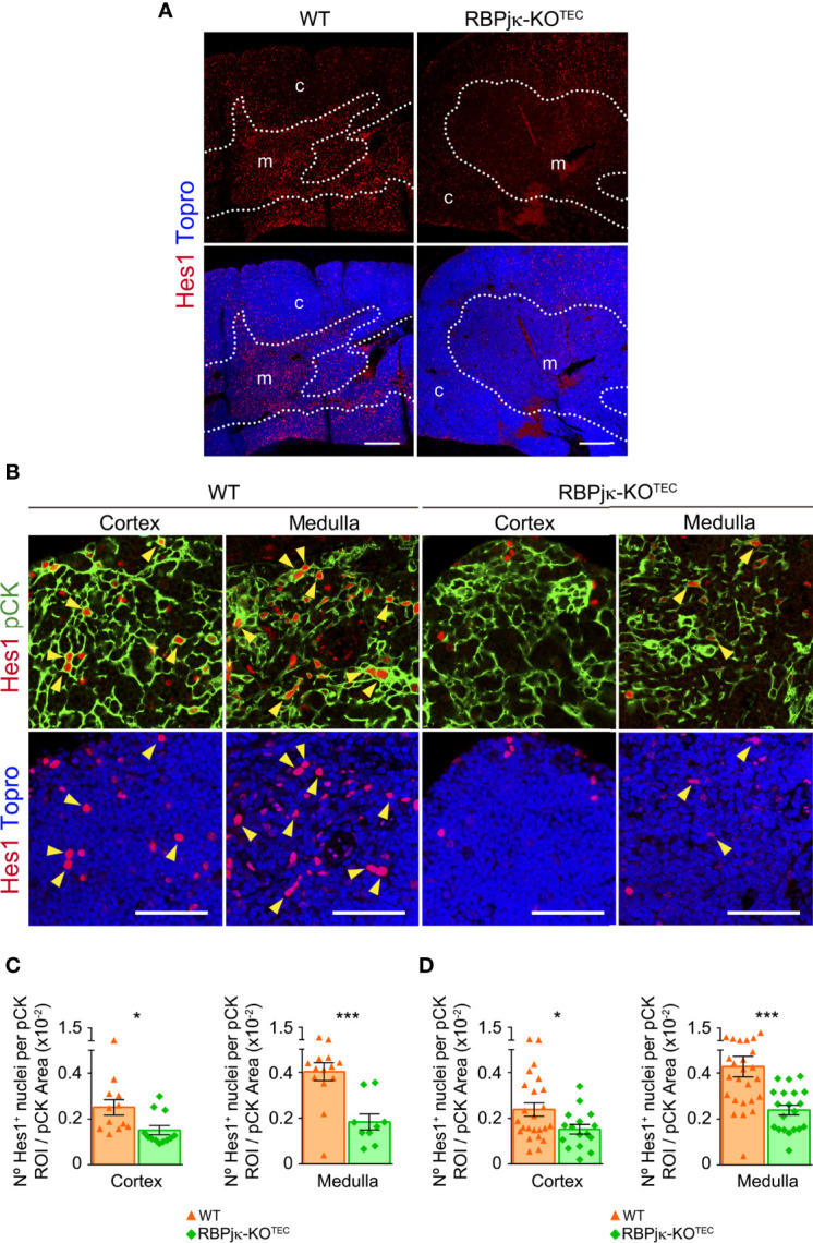
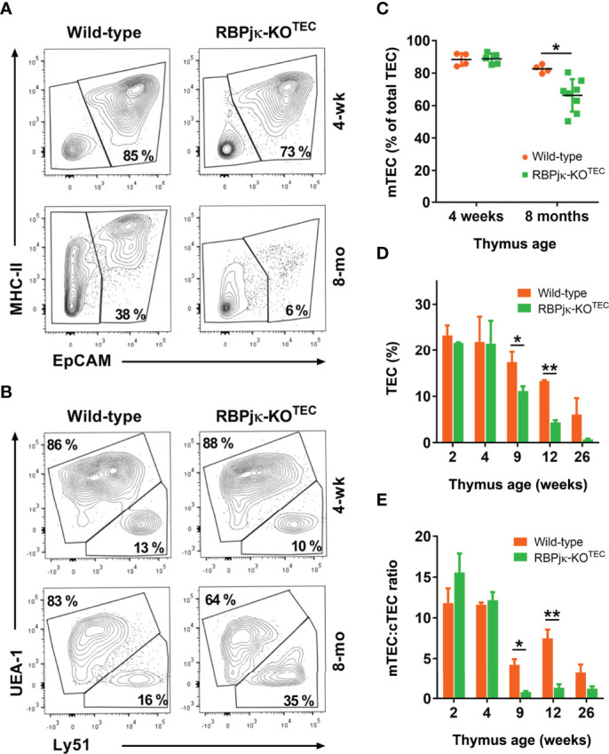
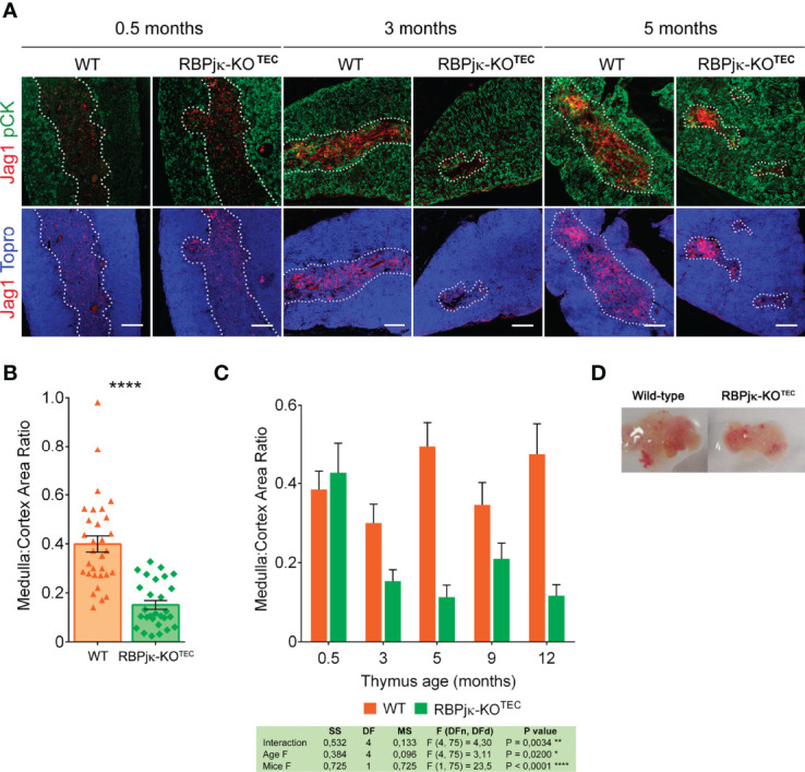
Notch signaling is crucial for fate specification and maturation of thymus-seeding progenitors along the T-cell lineage. Recent studies have extended the role of Notch signaling to thymic epithelial cells (TECs), showing that Notch regulates TEC progenitor maintenance and emergence of medullary TECs (mTECs) in fetal thymopoiesis. Based on immunohistochemistry studies of spatiotemporal regulation of Notch activation in the postnatal thymus, we show that Notch activation is not confined to fetal TECs. Rather, Notch signaling, likely mediated through the Notch1 receptor, is induced in postnatal cortical and medullary TECs, and increases significantly with age in the latter, in both humans and mice, suggesting a conserved role for Notch signaling in TEC homeostasis during thymus aging. To investigate the functional impact of Notch activation in postnatal TEC biology, we used a mouse model in which RPBJκ, the transcriptional effector of canonical Notch signaling, is deleted in epithelial cells, including TECs, under the control of the transcription factor Foxn1. Immunohistochemistry and flow cytometry analyses revealed no significant differences in TEC composition in mutant (RPBJκ-KO) and wild-type (WT) littermate mice at early postnatal ages. However, a significant reduction of the medullary region was observed in mutant compared to WT older thymi, which was accompanied by an accelerated decrease of postnatal mTEC numbers. Also, we found that organization and integrity of the postnatal thymic medulla critically depends on activation of the canonical Notch signaling pathway, as abrogation of Notch signaling in TECs led to the disruption of the medullary thymic microenvironment and to an accelerated thymus atrophy. These features paralleled a significant increase in the proportion of intrathymic non-T lineage cells, mostly B cells, and a slight decrease of DP thymocyte numbers compatible with a compromised thymic function in mutant mice. Therefore, impaired Notch signaling induced in embryonic development impacts postnatal TECs and leads to an accelerated mTEC degeneration and a premature thymus involution. Collectively, our data have uncovered a new role for Notch1 signaling in the control of adult mTEC homeostasis, and point toward Notch signaling manipulation as a novel strategy for thymus regeneration and functional recovery from immunosenescence.
Notch 信号通路对于胸腺祖细胞沿着 T 细胞谱系的命运特化和成熟至关重要。最近的研究将 Notch 信号通路的作用扩展到了胸腺上皮细胞 (TECs),表明 Notch 调节 TEC 祖细胞的维持和胎儿胸腺中髓质 TEC (mTEC)的出现。基于对出生后胸腺中 Notch 激活的时空调节的免疫组织化学研究,我们表明 Notch 激活不仅局限于胎儿 TECs。相反,Notch 信号通路,可能通过 Notch1 受体介导,在出生后的皮质和髓质 TEC 中被诱导,并且在后者中随着年龄的增长而显著增加,在人和小鼠中均如此,这表明 Notch 信号通路在胸腺衰老过程中对 TEC 稳态具有保守作用。为了研究 Notch 激活在出生后 TEC 生物学中的功能影响,我们使用了一种小鼠模型,其中 RPBJκ,即 Notch 信号通路的转录效应因子,在 Foxn1 转录因子的控制下在包括 TECs 在内的上皮细胞中被删除。免疫组织化学和流式细胞术分析显示,在早期出生后年龄,突变型 (RPBJκ-KO) 和野生型 (WT) 同窝仔鼠的 TEC 组成没有显著差异。然而,与 WT 较老的胸腺相比,突变型的髓质区域显著减小,这伴随着出生后 mTEC 数量的加速减少。此外,我们发现,出生后胸腺髓质的组织和完整性严重依赖于经典 Notch 信号通路的激活,因为 TEC 中的 Notch 信号通路的阻断导致了髓质胸腺微环境的破坏,并加速了胸腺的萎缩。这些特征与胸腺内非 T 谱系细胞(主要是 B 细胞)的比例显著增加以及 DP 胸腺细胞数量的轻微减少相一致,这与突变型小鼠的胸腺功能受损相符。因此,在胚胎发育过程中受损的 Notch 信号通路会影响出生后的 TEC,并导致 mTEC 的加速退化和胸腺的过早萎缩。总之,我们的数据揭示了 Notch1 信号通路在控制成人 mTEC 稳态中的新作用,并指出 Notch 信号通路的操纵可能是一种新的策略,用于胸腺再生和从免疫衰老中恢复功能。