• 文献检索
  • 文档翻译
  • 深度研究
  • 学术资讯
  • Suppr Zotero 插件Zotero 插件
  • 邀请有礼
  • 套餐&价格
  • 历史记录
应用&插件
Suppr Zotero 插件Zotero 插件浏览器插件Mac 客户端Windows 客户端微信小程序
定价
高级版会员购买积分包购买API积分包
服务
文献检索文档翻译深度研究API 文档MCP 服务
关于我们
关于 Suppr公司介绍联系我们用户协议隐私条款
关注我们

Suppr 超能文献

核心技术专利:CN118964589B侵权必究
粤ICP备2023148730 号-1Suppr @ 2026

文献检索

告别复杂PubMed语法,用中文像聊天一样搜索,搜遍4000万医学文献。AI智能推荐,让科研检索更轻松。

立即免费搜索

文件翻译

保留排版,准确专业,支持PDF/Word/PPT等文件格式,支持 12+语言互译。

免费翻译文档

深度研究

AI帮你快速写综述,25分钟生成高质量综述,智能提取关键信息,辅助科研写作。

立即免费体验

利用 micro-CT 通过对骨小梁内部形态的 3D 测量来判断已知创伤后时期的颅后骨折的年龄。

Utility of micro-CT for dating post-cranial fractures of known post-traumatic ages through 3D measurements of the trabecular inner morphology.

机构信息

Legal Medicine and Toxicology Unit, Department of Cardio-Thoraco-Vascular Sciences and Public Health, University of Padua, Padua, Italy.

Unit of Forensic Medicine, Department of Diagnostics and Public Health, University of Verona, P.le Scuro, 10, 37134, Verona, Italy.

出版信息

Sci Rep. 2022 Jun 22;12(1):10543. doi: 10.1038/s41598-022-14530-1.

DOI:10.1038/s41598-022-14530-1
PMID:35732857
原文链接:https://pmc.ncbi.nlm.nih.gov/articles/PMC9218115/
Abstract

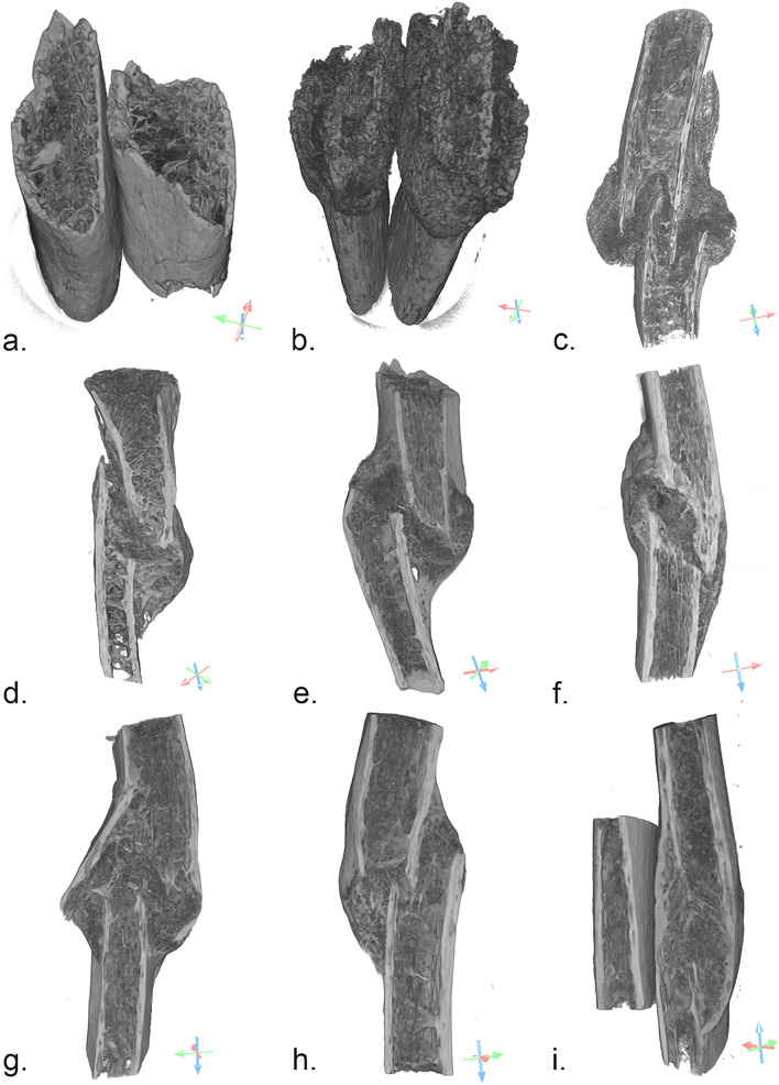
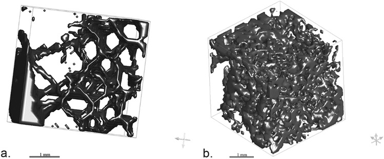
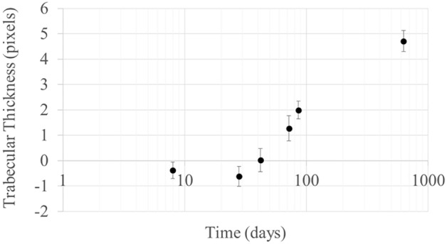
Fracture dating is an issue at the forefront of forensic sciences. While dating fracture is crucial to understanding and verifying the chronology of events in cases of abuse and violent death, its application is the subject of considerable discussion in the scientific community, filled with limitations and difficulties. Current methods for fracture dating are mainly based on a qualitative assessment through macroscopy, microscopy, and imaging and subject to variations depending on the experience of the observer. In this paper, we investigated the potential of quantifiable micro-CT analysis for fracture dating. Five histomorphometric parameters commonly used for the study of the 3D bone trabecular microarchitecture with micro-CT were calculated based on nine fractures of known post-traumatic ages, including the degree of anisotropy, connectivity density, bone volume fraction, trabecular thickness, and trabecular separation. As a result, trends in the evolution of the microarchitecture of the bone relative to age of the callus could be identified, in particular concerning anisotropy, trabecular separation and connectivity density, consistent with the healing bone process. The findings obtained in this pilot study encourage further research in quantifiable parameters of the bone microarchitecture as they could represent useful features for the construction of objective models for fracture dating.

摘要

骨折时间推断是法医学领域的一个前沿问题。虽然确定骨折时间对于了解和验证虐待和暴力死亡案件中事件的时间顺序至关重要,但在科学界,其应用存在相当大的争议,充满了局限性和困难。目前的骨折时间推断方法主要基于宏观、微观和影像学的定性评估,并且受观察者经验的影响。在本文中,我们研究了可量化的微 CT 分析在骨折时间推断中的潜力。基于 9 个已知创伤后年龄的骨折,计算了通常用于微 CT 研究三维骨小梁微结构的 5 个组织形态计量学参数,包括各向异性程度、连通密度、骨体积分数、骨小梁厚度和骨小梁分离。结果表明,相对于骨痂的年龄,骨微结构的演化趋势可以被识别出来,特别是各向异性、骨小梁分离和连通密度,这与愈合骨过程一致。这项初步研究的结果鼓励进一步研究骨微结构的可量化参数,因为它们可能是构建骨折时间推断的客观模型的有用特征。

https://cdn.ncbi.nlm.nih.gov/pmc/blobs/a099/9218115/eaa7c0cbea33/41598_2022_14530_Fig5_HTML.jpg
https://cdn.ncbi.nlm.nih.gov/pmc/blobs/a099/9218115/fd1fd3eeb727/41598_2022_14530_Fig1_HTML.jpg
https://cdn.ncbi.nlm.nih.gov/pmc/blobs/a099/9218115/2e6c14592382/41598_2022_14530_Fig2_HTML.jpg
https://cdn.ncbi.nlm.nih.gov/pmc/blobs/a099/9218115/77359be9ddbc/41598_2022_14530_Fig3_HTML.jpg
https://cdn.ncbi.nlm.nih.gov/pmc/blobs/a099/9218115/354da30e5670/41598_2022_14530_Fig4_HTML.jpg
https://cdn.ncbi.nlm.nih.gov/pmc/blobs/a099/9218115/eaa7c0cbea33/41598_2022_14530_Fig5_HTML.jpg

相似文献

1
Utility of micro-CT for dating post-cranial fractures of known post-traumatic ages through 3D measurements of the trabecular inner morphology.利用 micro-CT 通过对骨小梁内部形态的 3D 测量来判断已知创伤后时期的颅后骨折的年龄。
Sci Rep. 2022 Jun 22;12(1):10543. doi: 10.1038/s41598-022-14530-1.
2
The potential of micro-CT for dating post-cranial bone fractures: a macroscopic, radiographic, and microtomography study of fractures of known post-traumatic ages.微计算机断层扫描在鉴定颅外骨骨折时间上的潜力:已知创伤后年龄的骨折的宏观、放射学和微断层扫描研究。
Int J Legal Med. 2021 Sep;135(5):1913-1921. doi: 10.1007/s00414-021-02582-3. Epub 2021 Mar 27.
3
Micro-structural basis for particular vulnerability of the superolateral neck trabecular bone in the postmenopausal women with hip fractures.绝经后髋部骨折妇女超外侧颈骨小梁骨特殊脆弱性的微观结构基础。
Bone. 2012 Jan;50(1):63-8. doi: 10.1016/j.bone.2011.09.044. Epub 2011 Sep 22.
4
TBS reflects trabecular microarchitecture in premenopausal women and men with idiopathic osteoporosis and low-traumatic fractures.小梁骨评分(TBS)反映了绝经前患有特发性骨质疏松症和低创伤性骨折的女性和男性的小梁微结构。
Bone. 2015 Oct;79:259-66. doi: 10.1016/j.bone.2015.06.007. Epub 2015 Jun 16.
5
Microstructure of the hyoid bone based on micro-computed tomography findings.基于微计算机断层扫描结果的舌骨微观结构。
Medicine (Baltimore). 2020 Oct 30;99(44):e22246. doi: 10.1097/MD.0000000000022246.
6
Type 2 Diabetes Mellitus Increases the Risk to Hip Fracture in Postmenopausal Osteoporosis by Deteriorating the Trabecular Bone Microarchitecture and Bone Mass.2 型糖尿病通过破坏小梁骨微结构和骨量增加绝经后骨质疏松性髋部骨折的风险。
J Diabetes Res. 2019 Nov 7;2019:3876957. doi: 10.1155/2019/3876957. eCollection 2019.
7
Zoledronic acid suppresses callus remodeling but enhances callus strength in an osteoporotic rat model of fracture healing.唑来膦酸抑制骨质疏松性骨折愈合大鼠模型中骨痂的重塑,但增强了骨痂强度。
Bone. 2015 Dec;81:702-711. doi: 10.1016/j.bone.2015.09.018. Epub 2015 Oct 3.
8
Assessment of the healing of conservatively-treated scaphoid fractures using HR-pQCT.使用高分辨率外周定量CT评估保守治疗的舟骨骨折的愈合情况。
Bone. 2021 Dec;153:116161. doi: 10.1016/j.bone.2021.116161. Epub 2021 Aug 27.
9
Increases in BMD correlate with improvements in bone microarchitecture with teriparatide treatment in postmenopausal women with osteoporosis.在患有骨质疏松症的绝经后女性中,使用特立帕肽治疗后,骨密度的增加与骨微结构的改善相关。
J Bone Miner Res. 2007 Aug;22(8):1173-80. doi: 10.1359/jbmr.070413.
10
Micro-CT evaluation of bone defects: applications to osteolytic bone metastases, bone cysts, and fracture.微 CT 评估骨缺损:在溶骨性骨转移、骨囊肿和骨折中的应用。
Med Eng Phys. 2013 Nov;35(11):1645-50. doi: 10.1016/j.medengphy.2013.05.016. Epub 2013 Jul 3.

引用本文的文献

1
Radiomic analysis of postmortem lung changes: a PMCT-based approach for estimating the postmortem interval.尸检肺脏变化的影像组学分析:一种基于PMCT估计死后间隔时间的方法。
Forensic Sci Med Pathol. 2025 Aug 27. doi: 10.1007/s12024-025-01071-y.
2
Digital removal of dermal denticle layer using geometric AI from 3D CT scans of shark craniofacial structures enhances anatomical precision.利用几何人工智能从鲨鱼颅面结构的三维CT扫描中对真皮小齿层进行数字去除,可提高解剖精度。
Sci Rep. 2025 Jun 4;15(1):19659. doi: 10.1038/s41598-025-04442-1.
3
Imaging of musculoskeletal injury: timing estimation and medico-legal issues.

本文引用的文献

1
The potential of micro-CT for dating post-cranial bone fractures: a macroscopic, radiographic, and microtomography study of fractures of known post-traumatic ages.微计算机断层扫描在鉴定颅外骨骨折时间上的潜力:已知创伤后年龄的骨折的宏观、放射学和微断层扫描研究。
Int J Legal Med. 2021 Sep;135(5):1913-1921. doi: 10.1007/s00414-021-02582-3. Epub 2021 Mar 27.
2
Skull fractures in forensic putrefied/skeletonised cases: The challenge of estimating the post-traumatic interval.法医腐败/骨骼案例中的颅骨骨折:估计创伤后时间间隔的挑战。
Morphologie. 2020 Mar;104(344):27-37. doi: 10.1016/j.morpho.2020.01.002. Epub 2020 Feb 8.
3
肌肉骨骼损伤的影像学检查:时间估计及法医学问题
Radiol Med. 2025 Mar 28. doi: 10.1007/s11547-025-01990-4.
4
Targeting Optimal Bone Regions: Correlations Between Bone Density and DNA Quality in Small Skeletal Elements.靶向最佳骨区域:小骨骼元素中骨密度与DNA质量之间的相关性
Genes (Basel). 2025 Feb 27;16(3):291. doi: 10.3390/genes16030291.
5
Study of distribution and morphological characteristics of the trabecular bone in the uncinate process of the cervical spine using micro-computed tomography.使用显微计算机断层扫描对颈椎钩突内小梁骨的分布及形态特征进行研究。
PLoS One. 2025 Jan 3;20(1):e0315640. doi: 10.1371/journal.pone.0315640. eCollection 2025.
6
Effect of different running protocols on bone morphology and microarchitecture of the forelimbs in a male Wistar rat model.不同跑步方案对雄性 Wistar 大鼠前肢骨形态和微结构的影响。
PLoS One. 2024 Nov 7;19(11):e0308974. doi: 10.1371/journal.pone.0308974. eCollection 2024.
7
Determining rib fracture age from CT scans with a radiomics-based combined model: a multicenter retrospective study.基于影像组学的联合模型从CT扫描确定肋骨骨折年龄:一项多中心回顾性研究
Insights Imaging. 2023 Dec 10;14(1):214. doi: 10.1186/s13244-023-01546-y.
8
Osteoimmunology: The effect of autoimmunity on fracture healing and skeletal analysis.骨免疫学:自身免疫对骨折愈合及骨骼分析的影响
Forensic Sci Int Synerg. 2023 Mar 17;6:100326. doi: 10.1016/j.fsisyn.2023.100326. eCollection 2023.
9
Influence of Chinese Herbal Formula on Bone Characteristics of Cobb Broiler Chickens.中药配方对科布肉鸡骨骼特性的影响。
Genes (Basel). 2022 Oct 15;13(10):1865. doi: 10.3390/genes13101865.
Woven bone overview: structural classification based on its integral role in developmental, repair and pathological bone formation throughout vertebrate groups.
编织骨概述:根据其在整个脊椎动物群的发育、修复和病理性骨形成中的整体作用进行的结构分类。
Eur Cell Mater. 2019 Oct 1;38:137-167. doi: 10.22203/eCM.v038a11.
4
Histologic and radiological analysis on bone fractures: Estimation of posttraumatic survival time in skeletal trauma.骨创伤的组织学和影像学分析:骨创伤后存活时间的估计。
Forensic Sci Int. 2019 Sep;302:109909. doi: 10.1016/j.forsciint.2019.109909. Epub 2019 Aug 1.
5
Exiting the limbo of perimortem trauma: A brief review of microscopic markers of hemorrhaging and early healing signs in bone.走出濒死创伤的混沌:对骨内出血的微观标志物和早期愈合迹象的简要综述。
Forensic Sci Int. 2019 Sep;302:109856. doi: 10.1016/j.forsciint.2019.06.014. Epub 2019 Jun 19.
6
Forensic Radiology: A Primer.法医学放射学:入门指南。
Acad Radiol. 2019 Jun;26(6):820-830. doi: 10.1016/j.acra.2019.03.006. Epub 2019 Apr 17.
7
Histological ageing of fractures in infants: a practical algorithm for assessing infants suspected of accidental or non-accidental injury.婴儿骨折的组织学老化:评估疑似意外或非意外损伤婴儿的实用算法。
Histopathology. 2019 Jul;75(1):74-80. doi: 10.1111/his.13850. Epub 2019 Apr 29.
8
Recent advances in forensic anthropology.法医人类学的最新进展。
Forensic Sci Res. 2018 Jun 4;3(4):275-277. doi: 10.1080/20961790.2018.1466384. eCollection 2018.
9
Radiologic evaluation of fracture healing.骨折愈合的放射学评估
Skeletal Radiol. 2019 Mar;48(3):349-361. doi: 10.1007/s00256-018-3051-0. Epub 2018 Sep 21.
10
The detection of significant fractures in suspected infant abuse.疑似虐待婴儿中明显骨折的检测。
J Forensic Leg Med. 2018 Nov;60:9-14. doi: 10.1016/j.jflm.2018.09.002. Epub 2018 Sep 5.