Department of Joint Surgery, Center for Orthopaedic Surgery, The Third Affiliated Hospital of Southern Medical University, Guangzhou, China.
Department of Orthopedics, Orthopedic Hospital of Guangdong Province, Academy of Orthopedics·Guangdong Province, The Third Affiliated Hospital of Southern Medical University, Guangzhou, China.
Cell Death Dis. 2022 Jun 24;13(6):567. doi: 10.1038/s41419-022-04962-y.
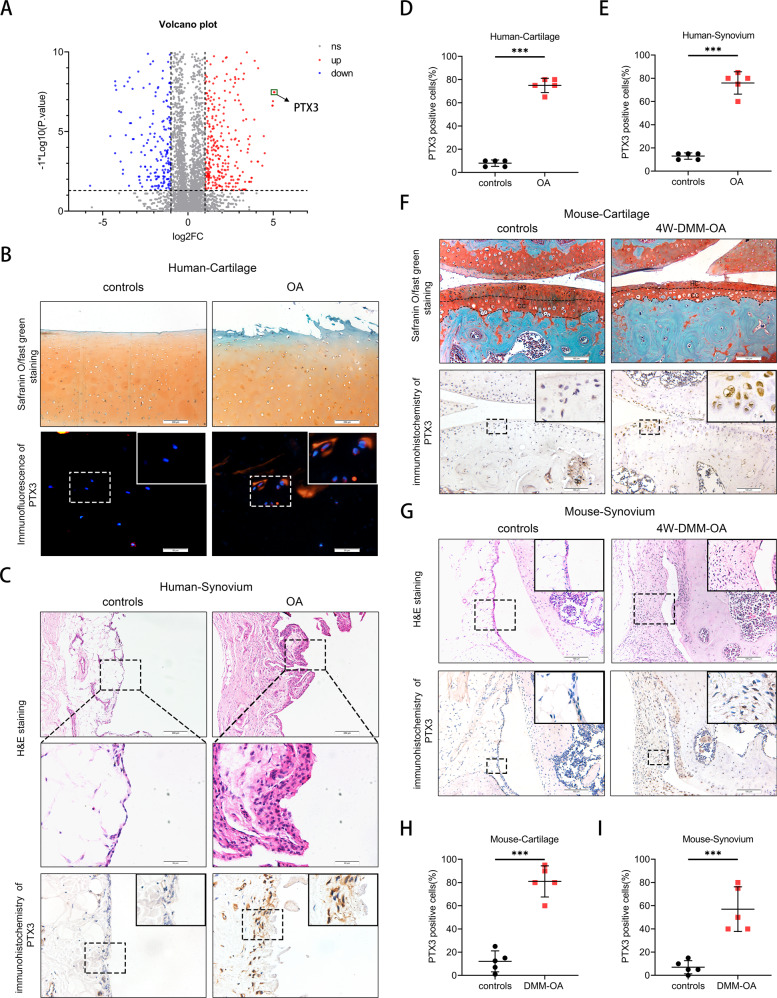
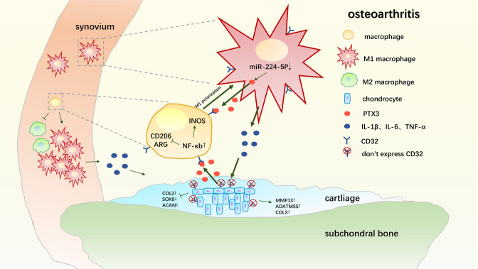
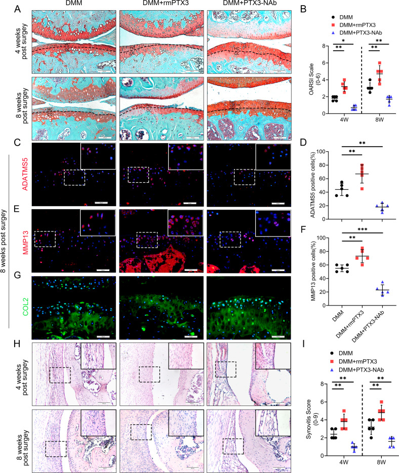
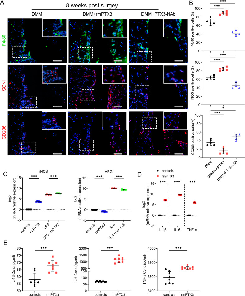
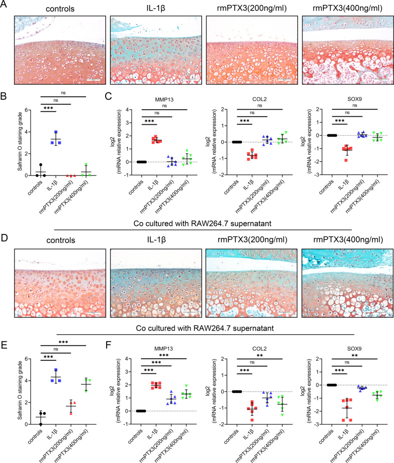
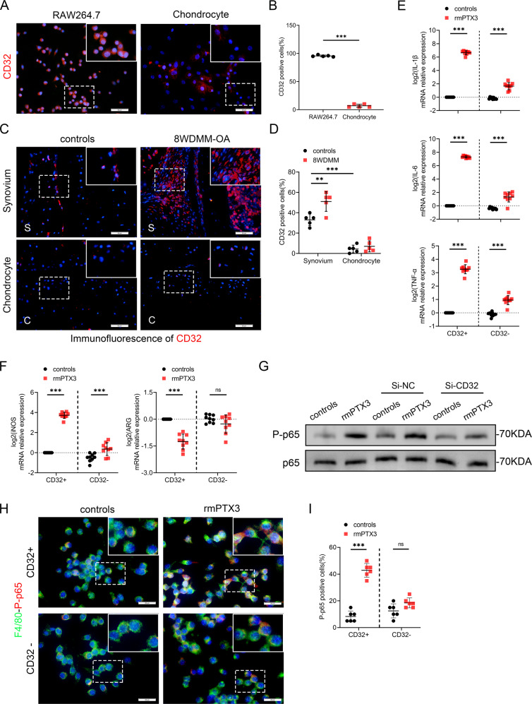
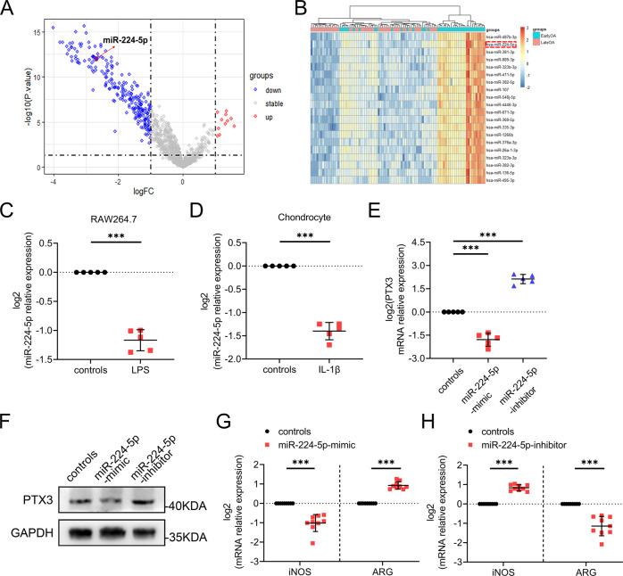
Emerging evidence has shown an imbalance in M1/M2 macrophage polarization to play an essential role in osteoarthritis (OA) progression. However, the underlying mechanistic basis for this polarization is unknown. RNA sequencing of OA M1-polarized macrophages found highly expressed levels of pentraxin 3 (PTX3), suggesting a role for PTX3 in OA occurrence and development. Herein, PTX3 was found to be increased in the synovium and articular cartilage of OA patients and OA mice. Intra-articular injection of PTX3 aggravated, while PTX3 neutralization reversed synovitis and cartilage degeneration. No metabolic disorder or proteoglycan loss were observed in cartilage explants when treated with PTX3 alone. However, cartilage explants exhibited an OA phenotype when treated with culture supernatants of macrophages stimulated with PTX3, suggesting that PTX3 did not have a direct effect on chondrocytes. Therefore, the OA anti-chondrogenic effects of PTX3 are primarily mediated through macrophages. Mechanistically, PTX3 was upregulated by miR-224-5p deficiency, which activated the p65/NF-κB pathway to promote M1 macrophage polarization by targeting CD32. CD32 was expressed by macrophages, that when stimulated with PTX3, secreted abundant pro-inflammation cytokines that induced severe articular cartilage damage. The paracrine interaction between macrophages and chondrocytes produced a feedback loop that enhanced synovitis and cartilage damage. The findings of this study identified a functional pathway important to OA development. Blockade of this pathway and PTX3 may prevent and treat OA.
新出现的证据表明,M1/M2 巨噬细胞极化失衡在骨关节炎(OA)进展中起着至关重要的作用。然而,这种极化的潜在机制基础尚不清楚。对 OA M1 极化巨噬细胞的 RNA 测序发现五聚素 3(PTX3)表达水平升高,提示 PTX3 在 OA 的发生和发展中起作用。本研究发现,OA 患者和 OA 小鼠的滑膜和关节软骨中 PTX3 增加。关节内注射 PTX3 加重了滑膜炎和软骨退化,而 PTX3 中和则逆转了滑膜炎和软骨退化。当单独用 PTX3 处理软骨外植体时,没有观察到代谢紊乱或蛋白聚糖丢失。然而,当用 PTX3 刺激的巨噬细胞培养上清处理软骨外植体时,软骨外植体表现出 OA 表型,表明 PTX3 对软骨细胞没有直接影响。因此,PTX3 对软骨的 OA 抗软骨作用主要是通过巨噬细胞介导的。在机制上,PTX3 被 miR-224-5p 缺陷上调,通过靶向 CD32 激活 p65/NF-κB 通路促进 M1 巨噬细胞极化。CD32 表达于巨噬细胞,当受到 PTX3 刺激时,分泌大量促炎细胞因子,导致严重的关节软骨损伤。巨噬细胞和软骨细胞之间的旁分泌相互作用产生了一个反馈环,增强了滑膜炎和软骨损伤。本研究的发现确定了一个对 OA 发展很重要的功能途径。阻断该途径和 PTX3 可能预防和治疗 OA。