Department of Biological Science, Center for Applied Biotechnology Studies, and Center for Computational and Applied Mathematics, College of Natural Sciences and Mathematics, California State University Fullerton, Fullerton, CA 92834, USA.
Department of Medicinal Chemistry and Molecular Pharmacology and the Purdue University Cancer Center, Purdue University, West Lafayette, IN 47907, USA.
Biomolecules. 2022 Jun 20;12(6):856. doi: 10.3390/biom12060856.
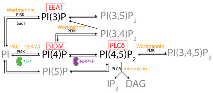
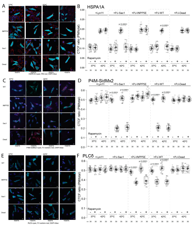
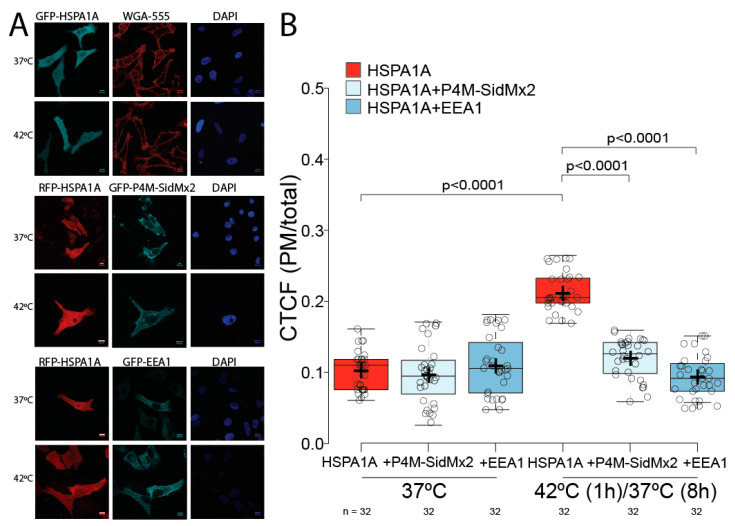
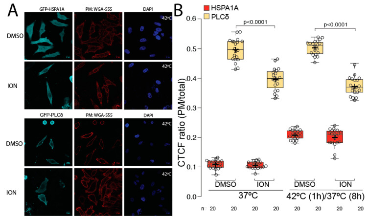
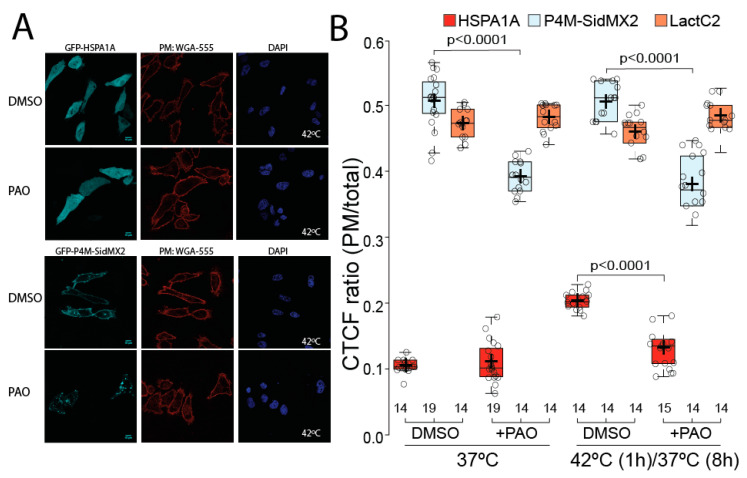
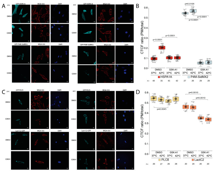
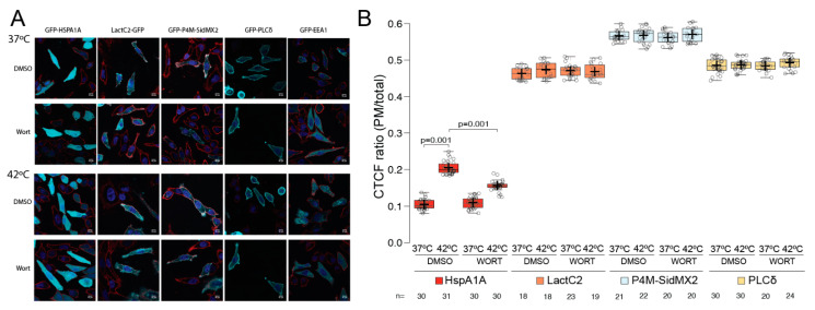
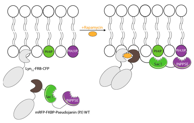
HSPA1A is a molecular chaperone that regulates the survival of stressed and cancer cells. In addition to its cytosolic pro-survival functions, HSPA1A also localizes and embeds in the plasma membrane (PM) of stressed and tumor cells. Membrane-associated HSPA1A exerts immunomodulatory functions and renders tumors resistant to standard therapies. Therefore, understanding and manipulating HSPA1A's surface presentation is a promising therapeutic. However, HSPA1A's pathway to the cell surface remains enigmatic because this protein lacks known membrane localization signals. Considering that HSPA1A binds to lipids, like phosphatidylserine (PS) and monophosphorylated phosphoinositides (PIPs), we hypothesized that this interaction regulates HSPA1A's PM localization and anchorage. To test this hypothesis, we subjected human cell lines to heat shock, depleted specific lipid targets, and quantified HSPA1A's PM localization using confocal microscopy and cell surface biotinylation. These experiments revealed that co-transfection of HSPA1A with lipid-biosensors masking PI(4)P and PI(3)P significantly reduced HSPA1A's heat-induced surface presentation. Next, we manipulated the cellular lipid content using ionomycin, phenyl arsine oxide (PAO), GSK-A1, and wortmannin. These experiments revealed that HSPA1A's PM localization was unaffected by ionomycin but was significantly reduced by PAO, GSK-A1, and wortmannin, corroborating the findings obtained by the co-transfection experiments. We verified these results by selectively depleting PI(4)P and PI(4,5)P using a rapamycin-induced phosphatase system. Our findings strongly support the notion that HSPA1A's surface presentation is a multifaceted lipid-driven phenomenon controlled by the binding of the chaperone to specific endosomal and PM lipids.
HSPA1A 是一种分子伴侣,可调节应激和癌细胞的存活。除了其细胞溶质的生存促进功能外,HSPA1A 还定位于应激和肿瘤细胞的质膜 (PM) 并嵌入其中。膜相关的 HSPA1A 发挥免疫调节功能,使肿瘤对标准疗法产生抗性。因此,了解和操纵 HSPA1A 的表面呈现是一种很有前途的治疗方法。然而,HSPA1A 到达细胞表面的途径仍然是个谜,因为这种蛋白质缺乏已知的膜定位信号。考虑到 HSPA1A 与脂质(如磷脂酰丝氨酸 (PS) 和单磷酸化的磷酸肌醇 (PIPs))结合,我们假设这种相互作用调节 HSPA1A 的 PM 定位和锚定。为了验证这一假设,我们使人类细胞系经受热休克,耗尽特定的脂质靶点,并使用共聚焦显微镜和细胞表面生物素化来定量 HSPA1A 的 PM 定位。这些实验表明,HSPA1A 与掩盖 PI(4)P 和 PI(3)P 的脂质生物传感器共转染显着降低了 HSPA1A 的热诱导表面呈现。接下来,我们使用离子霉素、苯砷氧化物 (PAO)、GSK-A1 和渥曼青霉素操纵细胞内脂质含量。这些实验表明,HSPA1A 的 PM 定位不受离子霉素影响,但被 PAO、GSK-A1 和渥曼青霉素显着降低,这与共转染实验的结果相符。我们通过使用雷帕霉素诱导的磷酸酶系统选择性耗尽 PI(4)P 和 PI(4,5)P 验证了这些结果。我们的研究结果强烈支持这样一种观点,即 HSPA1A 的表面呈现是一种多方面的脂质驱动现象,受伴侣蛋白与特定内体和 PM 脂质结合的控制。