Department of Circulation and Medical Imaging, Faculty of Medicine and Health Sciences, Norwegian University of Science and Technology, Trondheim, Norway.
Department of Radiology and Nuclear Medicine, St. Olavs Hospital, Trondheim University Hospital, Trondheim, Norway.
Neuroradiology. 2022 Dec;64(12):2217-2226. doi: 10.1007/s00234-022-02998-7. Epub 2022 Jun 27.
To assess the ability of 7 T MRI to detect hippocampal DWI lesions in the acute phase of TGA compared to 1.5 T/3 T MRI.
Patients with a clinical diagnosis consistent with TGA and a 1.5/3 T MRI underwent an additional 7 T MRI when the 7 T system was available for clinical use, thus serving as their own controls.
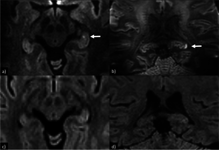
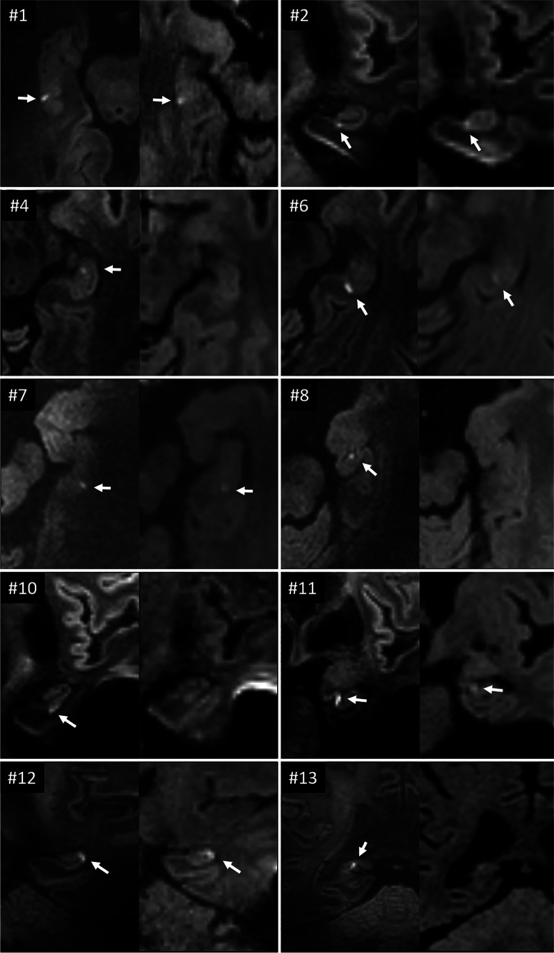
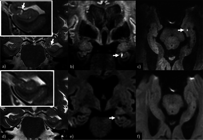
Thirteen TGA patients with a median age of 68.5 years (range 46-77 years) were included and imaged at 1.5/3 T (median 17 h after onset of symptoms, range 3-23 h) and 7 T (median 23 h after onset, range 15-46 h). The 7 T MRIs were performed a median of 15 h after the 1.5/3 T MRIs (range 1-28 h). At 1.5/3 T, six patients (46%) were found to have at least one hippocampal DWI-lesions supporting the TGA diagnosis, which increased to 11 patients (85%) when examined at 7 T (p = 0.03). At 1.5/3 T, nine hippocampal DWI lesions were detected, which increased to 19 at 7 T, giving an increased detection rate of 111% (p = 0.002). Both neuroradiologists found the hippocampal DWI lesions at 7 T to have higher conspicuity and be easier to categorize as true findings compared to 1.5/3 T.
Seven-Tesla MRI showed both a statistically significant increase in the total number of detected hippocampal DWI lesions and the proportion of patients with at least one hippocampal DWI lesion supporting the TGA diagnosis compared to 1.5/3 T. Clinical use of 7 T will increase the number of patients having their TGA diagnosis supported by MRI, which can be especially useful in patients with negative 1.5/3 T MRI and low clinical certainty.
评估 7T MRI 在急性 TGA 中检测海马弥散加权成像(DWI)病变的能力,并与 1.5T/3T MRI 进行比较。
当 7T 系统可供临床使用时,对符合 TGA 临床诊断且已行 1.5/3T MRI 检查的患者进行额外的 7T MRI 检查,这些患者作为自身对照。
共纳入 13 例 TGA 患者,中位年龄 68.5 岁(范围 46-77 岁),分别在 1.5/3T(中位发病后 17h,范围 3-23h)和 7T(中位发病后 23h,范围 15-46h)进行检查。7T MRI 检查在完成 1.5/3T MRI 检查后中位时间 15h(范围 1-28h)进行。在 1.5/3T 上,6 例(46%)患者至少有 1 个海马 DWI 病变支持 TGA 诊断,而在 7T 上增加到 11 例(85%)(p=0.03)。在 1.5/3T 上共发现 9 个海马 DWI 病变,在 7T 上增加到 19 个,检出率增加了 111%(p=0.002)。两位神经放射科医生均发现,与 1.5/3T 相比,7T 上的海马 DWI 病变更明显,更容易归类为真正的发现。
与 1.5/3T 相比,7T MRI 显示在检测到的总海马 DWI 病变数量以及至少有 1 个海马 DWI 病变支持 TGA 诊断的患者比例方面均有统计学显著增加。7T 的临床应用将增加 MRI 支持 TGA 诊断的患者数量,这在 1.5/3T MRI 阴性且临床确定性低的患者中尤其有用。