Boissy R E, Moellmann G E, Lerner A B
Am J Pathol. 1987 May;127(2):380-8.
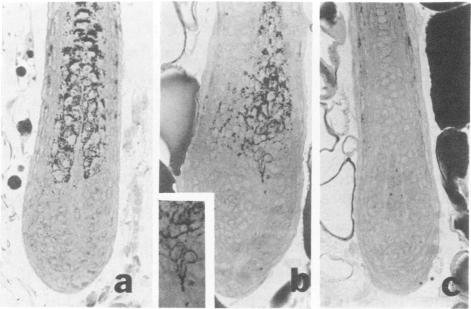
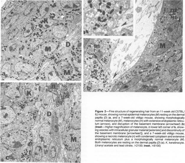
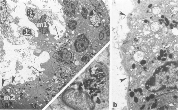
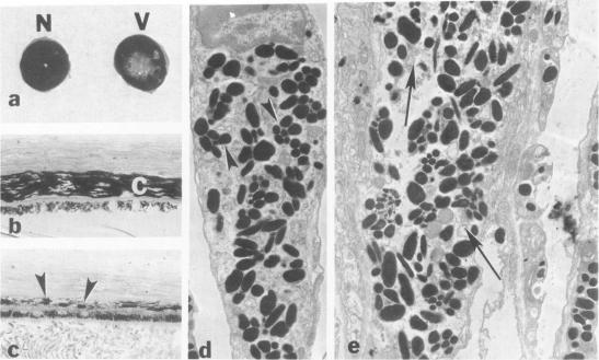
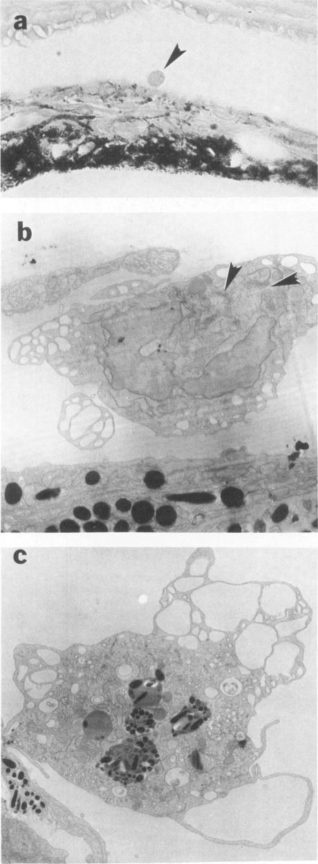
The vitiligo mouse C57BL/6J Ler-vit/vit is a new, murine model for vitiligo in humans. It was studied with respect to morphology and fine structure of melanocytes in hair and eyes before and during depigmentation. The coat of vitiligo mice lightens progressively with age because of an increase in the ratio of white to pigmented hairs with each molt. The bulbs of white hairs are devoid of pigment, and they lack melanocytes. In other respects the epithelium is morphologically normal as determined by light and electron microscopy. The bulbs of pigmented hairs are histologically normal. By electron microscopy, however, some of the melanocytes are shown to have undergone degenerative changes. In addition, disruption of the basement membrane underlying the melanocytes and herniation of melanocytes into dermal papillae were observed at various stages of hair growth. Papillary melanophages are prominent in pigmented as well as in white hair bulbs. Newborn vitiligo mice have no uveal pigment. Pigment appears in the iris and ciliary body by Day 4 and in the choroid by Week 3. On Day 4, along with pigmentation, conspicuous spherical amelanotic cells appear over the anterior border of the iris. These cells become numerous in the ensuing weeks and gradually acquire large melanophagosomes. They occur also in the stroma of the iris and the ciliary body, associated with necrotic melanocytes. The spherical cells are identical to the clump cells of Koganei and are far more numerous in vitiligo mice than in controls. Macroscopically, no progressive decrease in iridial pigment is apparent for the life of the vitiligo mouse. In the choroid, an amelanotic patch surrounds the optic nerve. In the pigmented areas, melanocytes show compartmentalization of melanosomes and degeneration. The retinal pigment epithelium generally appeared continuous. In older animals some epithelial cells contained large fat bodies or were devoid of melanin.
白癜风小鼠C57BL/6J Ler-vit/vit是一种新的人类白癜风小鼠模型。对其在色素脱失前和色素脱失期间毛发和眼睛中黑素细胞的形态和精细结构进行了研究。由于每次换毛时白毛与有色毛的比例增加,白癜风小鼠的皮毛会随着年龄的增长而逐渐变浅。白毛的毛囊没有色素,也没有黑素细胞。在其他方面,通过光学显微镜和电子显微镜观察,上皮在形态上是正常的。有色毛的毛囊在组织学上是正常的。然而,通过电子显微镜观察,一些黑素细胞显示出退行性变化。此外,在毛发生长的各个阶段都观察到黑素细胞下方的基底膜破坏以及黑素细胞疝入真皮乳头。乳头黑素巨噬细胞在有色毛囊和白色毛囊中都很突出。新生白癜风小鼠没有葡萄膜色素。第4天时色素出现在虹膜和睫状体中,第3周时出现在脉络膜中。在第4天,随着色素沉着,明显的球形无黑素细胞出现在虹膜的前缘。在接下来的几周里,这些细胞数量增多,并逐渐获得大的黑素吞噬体。它们也出现在虹膜和睫状体的基质中,与坏死的黑素细胞有关。这些球形细胞与小金井的团块细胞相同,在白癜风小鼠中比在对照小鼠中要多得多。宏观上,在白癜风小鼠的生命期内,虹膜色素没有明显的逐渐减少。在脉络膜中,视神经周围有一个无黑素斑。在色素沉着区域,黑素细胞显示出黑素小体的分隔和退化。视网膜色素上皮通常看起来是连续的。在老年动物中,一些上皮细胞含有大的脂肪体或没有黑色素。