• 文献检索
  • 文档翻译
  • 深度研究
  • 学术资讯
  • Suppr Zotero 插件Zotero 插件
  • 邀请有礼
  • 套餐&价格
  • 历史记录
应用&插件
Suppr Zotero 插件Zotero 插件浏览器插件Mac 客户端Windows 客户端微信小程序
定价
高级版会员购买积分包购买API积分包
服务
文献检索文档翻译深度研究API 文档MCP 服务
关于我们
关于 Suppr公司介绍联系我们用户协议隐私条款
关注我们

Suppr 超能文献

核心技术专利:CN118964589B侵权必究
粤ICP备2023148730 号-1Suppr @ 2026

文献检索

告别复杂PubMed语法,用中文像聊天一样搜索,搜遍4000万医学文献。AI智能推荐,让科研检索更轻松。

立即免费搜索

文件翻译

保留排版,准确专业,支持PDF/Word/PPT等文件格式,支持 12+语言互译。

免费翻译文档

深度研究

AI帮你快速写综述,25分钟生成高质量综述,智能提取关键信息,辅助科研写作。

立即免费体验

小儿阑尾外径的超声评估。

Ultrasonographic evaluation of cecal appendix diameter in pediatric population.

机构信息

Hospital Israelita Albert Einstein, São Paulo, SP, Brazil.

Hospital das Clínicas, Faculdade de Medicina, Universidade de São Paulo, São Paulo, SP, Brazil.

出版信息

Einstein (Sao Paulo). 2022 Jul 4;20:eAO6935. doi: 10.31744/einstein_journal/2022AO6935. eCollection 2022.

DOI:10.31744/einstein_journal/2022AO6935
PMID:35792758
原文链接:https://pmc.ncbi.nlm.nih.gov/articles/PMC9239537/
Abstract

OBJECTIVE

To stratify ultrasound samples in a pediatric population undergoing evaluation for acute appendicitis to examine the variability in cecal appendix diameter, in different age groups, and to determine whether there is a prevalent value for each age group.

METHODS

A retrospective cross-sectional study with 196 children aged 0 to 15 years. Data were extracted from reports of ultrasound examinations carried out between 2008 and 2015. Children with sonographic diagnosis of appendicitis or other signs of periappendiceal inflammation were excluded.

RESULTS

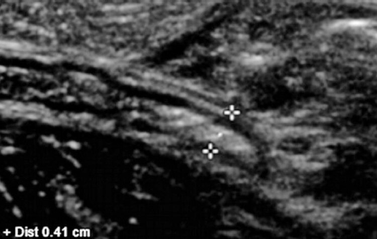
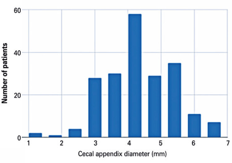
The evaluation of the anteroposterior measurement of the cecal appendix revealed a mean diameter of 4.14mm (standard deviation: 0.93mm; 95%CI: 3.86-4.14). Cecal appendix diameter did not differ significant between age groups.

CONCLUSION

Evaluation of the anteroposterior diameter of the cecal appendix in centimeters in a sample of 196 children aged 0 to15 years revealed a mean diameter of 4.14mm (standard deviation, 0.93mm. There were no significant differences in cecal appendix diameter following stratification by age. Results indicate a single value can be adopted for mean cecal appendix diameter in pediatric populations.

摘要

目的

对接受急性阑尾炎评估的儿科人群的超声样本进行分层,以检查不同年龄组盲肠阑尾直径的变化,并确定每个年龄组是否存在常见值。

方法

这是一项回顾性的病例对照研究,共纳入 196 名 0 至 15 岁的儿童。数据来自 2008 年至 2015 年进行的超声检查报告。排除了超声诊断为阑尾炎或其他阑尾周围炎症迹象的儿童。

结果

对盲肠阑尾前后径的评估显示平均直径为 4.14mm(标准差:0.93mm;95%CI:3.86-4.14)。不同年龄组之间盲肠阑尾直径无显著差异。

结论

对 196 名 0 至 15 岁儿童的盲肠阑尾前后径进行评估,结果显示平均直径为 4.14mm(标准差,0.93mm)。根据年龄分层,盲肠阑尾直径无显著差异。结果表明,在儿科人群中可以采用单个均值来表示盲肠阑尾直径。

https://cdn.ncbi.nlm.nih.gov/pmc/blobs/a8c0/9239537/162f5b364807/2317-6385-eins-20-eAO6935-gf05.jpg
https://cdn.ncbi.nlm.nih.gov/pmc/blobs/a8c0/9239537/30d9ce54fa17/2317-6385-eins-20-eAO6935-gf01.jpg
https://cdn.ncbi.nlm.nih.gov/pmc/blobs/a8c0/9239537/7c9d346e2537/2317-6385-eins-20-eAO6935-gf02.jpg
https://cdn.ncbi.nlm.nih.gov/pmc/blobs/a8c0/9239537/55ea0b35b538/2317-6385-eins-20-eAO6935-gf03.jpg
https://cdn.ncbi.nlm.nih.gov/pmc/blobs/a8c0/9239537/162f5b364807/2317-6385-eins-20-eAO6935-gf05.jpg

相似文献

1
Ultrasonographic evaluation of cecal appendix diameter in pediatric population.小儿阑尾外径的超声评估。
Einstein (Sao Paulo). 2022 Jul 4;20:eAO6935. doi: 10.31744/einstein_journal/2022AO6935. eCollection 2022.
2
Appendix not seen: the predictive value of secondary inflammatory sonographic signs.未见到阑尾:继发性炎症超声征象的预测价值。
Pediatr Emerg Care. 2013 Apr;29(4):435-9. doi: 10.1097/PEC.0b013e318289e8d5.
3
Independent predictors of acute appendicitis on CT with pathologic correlation.CT上急性阑尾炎的独立预测因素及其病理相关性
Acad Radiol. 2008 Aug;15(8):996-1003. doi: 10.1016/j.acra.2008.02.009.
4
Bedside ultrasonography as an adjunct to routine evaluation of acute appendicitis in the emergency department.床边超声检查作为急诊科急性阑尾炎常规评估的辅助手段。
West J Emerg Med. 2014 Nov;15(7):808-15. doi: 10.5811/westjem.2014.9.21491. Epub 2014 Sep 18.
5
Lymphoid Hyperplasia of the Appendix: A Potential Pitfall in the Sonographic Diagnosis of Appendicitis.阑尾淋巴样增生:超声诊断阑尾炎时的一个潜在陷阱。
AJR Am J Roentgenol. 2016 Jan;206(1):189-94. doi: 10.2214/AJR.15.14846.
6
Variation in imaging outcomes associated with individual sonographers and radiologists in pediatric acute appendicitis: a retrospective cohort of 9271 examinations.在儿科急性阑尾炎中,与个别超声医师和放射科医师相关的影像学结果的变化:一项对 9271 例检查的回顾性队列研究。
Eur Radiol. 2021 Nov;31(11):8565-8577. doi: 10.1007/s00330-021-07939-1. Epub 2021 Apr 21.
7
Cecal apical changes with appendicitis: diagnosing appendicitis when the appendix is borderline abnormal or not seen.阑尾炎时盲肠顶端的改变:当阑尾边界不清异常或未显示时诊断阑尾炎
J Comput Assist Tomogr. 1999 Jan-Feb;23(1):55-9. doi: 10.1097/00004728-199901000-00012.
8
Diagnostic Performance of US for Differentiating Perforated from Nonperforated Pediatric Appendicitis: A Prospective Cohort Study.超声对小儿穿孔性与非穿孔性阑尾炎的诊断性能:一项前瞻性队列研究。
Radiology. 2017 Mar;282(3):835-841. doi: 10.1148/radiol.2016160175. Epub 2016 Oct 31.
9
Appendiceal diameter: CT versus sonographic measurements.阑尾直径:CT与超声测量对比
Pediatr Radiol. 2016 Mar;46(3):316-21. doi: 10.1007/s00247-015-3491-9. Epub 2015 Nov 16.
10
Outcomes for Children With a Nonvisualized Appendix on Ultrasound.超声检查未显示阑尾的儿童的预后情况。
Pediatr Emerg Care. 2021 Aug 1;37(8):e456-e460. doi: 10.1097/PEC.0000000000001672.

引用本文的文献

1
In Vivo Measurements of Appendiceal Base - Implications on Pediatric Laparoscopic Appendectomy.阑尾根部的体内测量——对小儿腹腔镜阑尾切除术的影响
JSLS. 2024 Jul-Sep;28(3). doi: 10.4293/JSLS.2024.00033. Epub 2025 Jan 10.

本文引用的文献

1
Improving diagnostic accuracy in clinically ambiguous paediatric appendicitis: a retrospective review of ultrasound and pathology findings with focus on the non-visualised appendix.提高临床不明确性小儿阑尾炎的诊断准确性:超声与病理检查结果的回顾性分析,重点关注未可视化阑尾。
Br J Radiol. 2019 Jan;92(1093):20180585. doi: 10.1259/bjr.20180585. Epub 2018 Sep 12.
2
Development and validation of an ultrasound scoring system for children with suspected acute appendicitis.疑似急性阑尾炎儿童超声评分系统的开发与验证
Pediatr Radiol. 2015 Dec;45(13):1945-52. doi: 10.1007/s00247-015-3443-4. Epub 2015 Aug 18.
3
Pediatric appendiceal ultrasound: accuracy, determinacy and clinical outcomes.
小儿阑尾超声检查:准确性、确定性及临床结果
Pediatr Radiol. 2015 Dec;45(13):1934-44. doi: 10.1007/s00247-015-3432-7. Epub 2015 Aug 18.
4
Does appendiceal diameter change with age? A sonographic study.阑尾直径会随年龄变化吗?一项超声研究。
AJR Am J Roentgenol. 2014 Nov;203(5):1120-6. doi: 10.2214/AJR.13.12205.
5
RSNA centennial article: ALARA 1912: "As low a dose as possible" a century ago.RSNA百年纪念文章:1912年的合理可达到的最低辐射剂量:一个世纪前的“尽可能低的剂量”
Radiographics. 2014 Sep-Oct;34(5):1457-60. doi: 10.1148/rg.345130136.
6
Acute appendicitis: investigating an optimal outer appendiceal diameter cut-point in a pediatric population.急性阑尾炎:探究儿科人群中阑尾外径的最佳切点
J Emerg Med. 2014 Feb;46(2):157-64. doi: 10.1016/j.jemermed.2013.08.027. Epub 2013 Oct 7.
7
Changes in the length and diameter of the normal appendix throughout childhood.儿童期正常阑尾长度和直径的变化。
J Pediatr Surg. 2013 Jul;48(7):1535-9. doi: 10.1016/j.jpedsurg.2013.02.035.
8
Performance of ultrasound in the diagnosis of appendicitis in children in a multicenter cohort.超声在多中心队列中儿童阑尾炎诊断中的表现。
Acad Emerg Med. 2013 Jul;20(7):697-702. doi: 10.1111/acem.12161.
9
Reevaluating the sonographic criteria for acute appendicitis in children: a review of the literature and a retrospective analysis of 246 cases.重新评估儿童急性阑尾炎的超声标准:文献复习和 246 例回顾性分析。
Acad Radiol. 2012 Nov;19(11):1382-94. doi: 10.1016/j.acra.2012.06.014. Epub 2012 Sep 2.
10
Sonographic appearance of the normal appendix in children.儿童正常阑尾的超声表现。
J Clin Ultrasound. 2011 May;39(4):183-6. doi: 10.1002/jcu.20807. Epub 2011 Mar 21.