State Key Laboratory of Medicinal Chemical Biology, Frontiers Science Center for Cell Responses, College of Life Sciences, Nankai University, No. 94 Weijin Road, Tianjin, 300071, China.
Organ Transplant Department, Tianjin First Central Hospital, School of Medicine, Nankai University, Tianjin, China.
Cell Commun Signal. 2022 Jul 7;20(1):102. doi: 10.1186/s12964-022-00911-6.
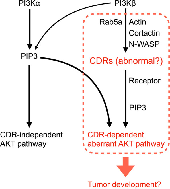
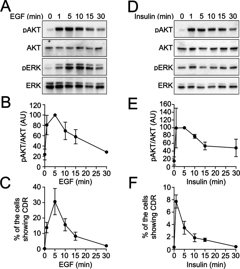
Circular dorsal ruffles (CDRs) are rounded membrane ruffles induced on the dorsal surfaces of cells stimulated by growth factors (GF). They can serve as signal platforms to activate AKT protein kinase. After GF stimulation, phosphatidylinositol 3-kinase (PI3K) generates phosphatidylinositol (3,4,5)-triphosphate (PIP3) in the plasma membrane. PIP3 accumulates inside CDRs, recruits AKT into the structures, and phosphorylates them (pAKT). Given the importance of the PI3K-AKT pathway in GF signaling, CDRs are likely involved in cell growth. Interestingly, some cancer cell lines express CDRs. We hypothesized that CDRs contribute to carcinogenesis by modulating the AKT pathway. In the present study, we identified CDR-expressing cancer cell lines and investigated their cellular functions.
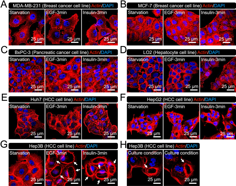
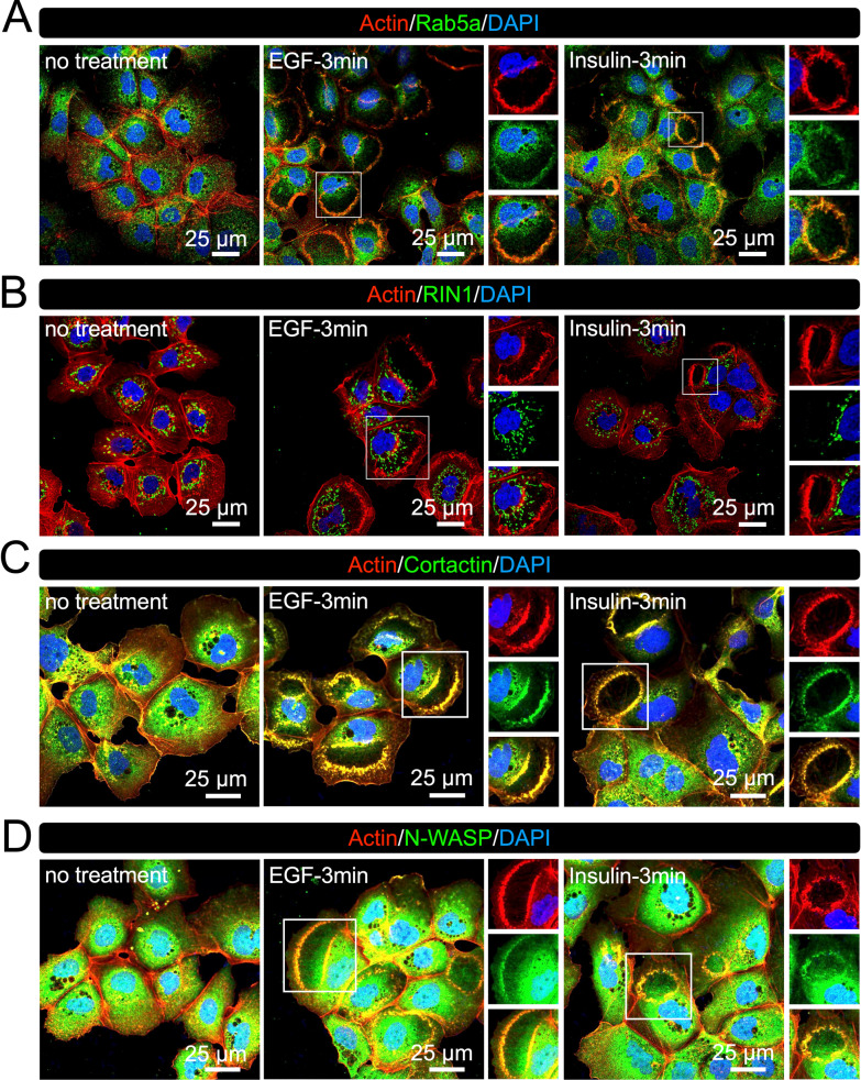
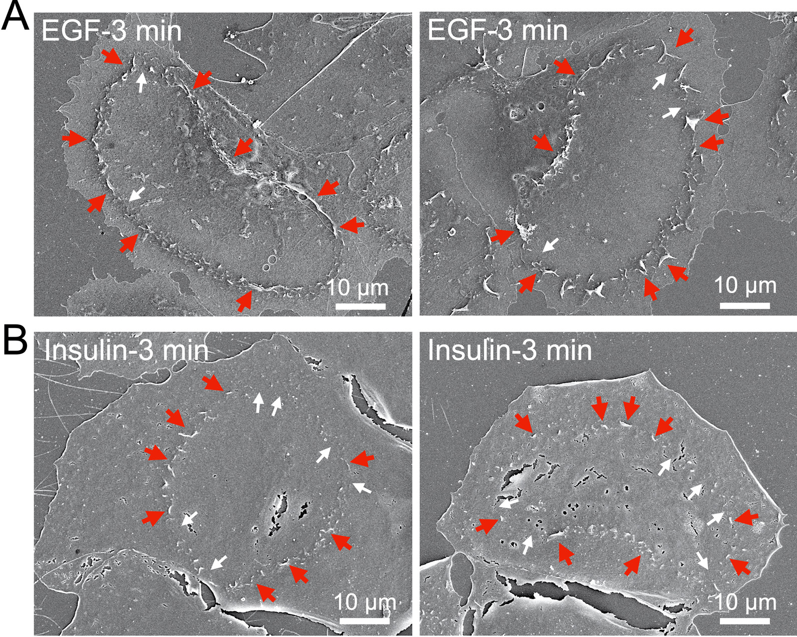
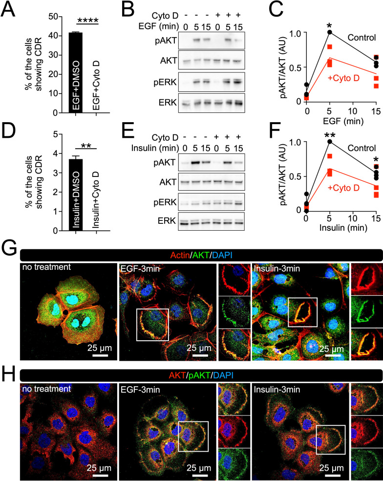
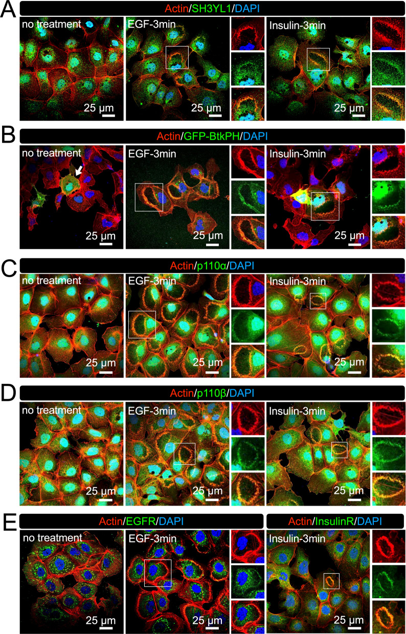
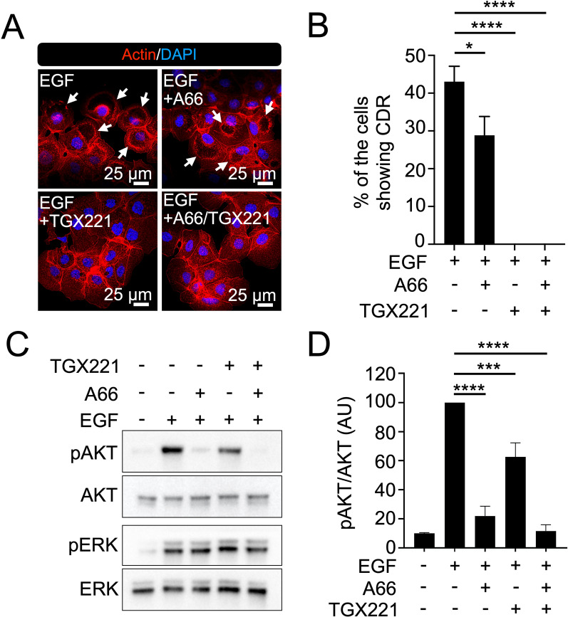
CDR formation was examined in six cancer cell lines in response to epidermal growth factor (EGF) and insulin. The morphology of the CDRs was characterized, and the related signaling molecules were observed using confocal and scanning electron microscopy. The role of CDRs in the AKT pathway was studied using biochemical analysis. The actin inhibitor cytochalasin D (Cyto D) and the PI3K inhibitor TGX221 were used to block CDRs.
GF treatment induced CDRs in the hepatocellular carcinoma (HCC) Hep3B cell line, but not in others, including HCC cell lines HepG2 and Huh7, and the LO2 hepatocyte cell line. Confocal microscopy and western blot analysis showed that the PI3K-PIP3-AKT pathway was activated at the CDRs and that receptor proteins were recruited to the structures. Cyto D and TGX221 completely blocked CDRs and partially attenuated GF-induced pAKT. These results indicate that CDRs regulate the receptor-mediated PI3K-AKT pathway in Hep3B cells and the existence of CDR-independent pAKT mechanisms.
Our results showed that CDRs modulate the AKT pathway in Hep3B cells. Since CDRs were not observed in other HCC and hepatocyte cell lines, we propose that CDRs in Hep3B would determine the carcinoma characteristic of the cell by aberrantly triggering the AKT pathway. Signaling molecules involved in CDR formation are promising therapeutic targets for some types of HCC. Video abstract.
圆形背侧皱襞 (CDR) 是细胞在生长因子 (GF) 刺激下诱导的圆形膜皱襞。它们可以作为激活 AKT 蛋白激酶的信号平台。GF 刺激后,磷脂酰肌醇 3-激酶 (PI3K) 在质膜中产生磷脂酰肌醇 (3,4,5)-三磷酸 (PIP3)。PIP3 在 CDR 内积聚,募集 AKT 进入结构,并使其磷酸化 (pAKT)。鉴于 PI3K-AKT 途径在 GF 信号转导中的重要性,CDR 可能参与细胞生长。有趣的是,一些癌细胞系表达 CDR。我们假设 CDR 通过调节 AKT 途径促进癌变。在本研究中,我们鉴定了表达 CDR 的癌细胞系,并研究了它们的细胞功能。
用表皮生长因子 (EGF) 和胰岛素检测 6 种癌细胞系中 CDR 的形成。用共聚焦和扫描电子显微镜观察 CDR 的形态,观察相关信号分子。用生化分析研究 CDR 在 AKT 途径中的作用。用肌动蛋白抑制剂细胞松弛素 D (Cyto D) 和 PI3K 抑制剂 TGX221 阻断 CDR。
GF 处理诱导肝癌 (HCC) Hep3B 细胞系中 CDR 的形成,但在其他细胞系中不诱导,包括 HCC 细胞系 HepG2 和 Huh7 以及 LO2 肝细胞系。共聚焦显微镜和 Western blot 分析表明,PI3K-PIP3-AKT 途径在 CDR 处被激活,受体蛋白被募集到结构中。Cyto D 和 TGX221 完全阻断 CDR,并部分减弱 GF 诱导的 pAKT。这些结果表明 CDR 调节 Hep3B 细胞中受体介导的 PI3K-AKT 途径,并且存在 CDR 独立的 pAKT 机制。
我们的结果表明,CDR 调节 Hep3B 细胞中的 AKT 途径。由于在其他 HCC 和肝细胞系中未观察到 CDR,我们提出 Hep3B 中的 CDR 通过异常触发 AKT 途径来决定细胞的癌特征。参与 CDR 形成的信号分子是某些类型 HCC 的有前途的治疗靶点。视频摘要。