Sala-Padro Jacint, Gifreu-Fraixino Ariadna, Miró Júlia, Rodriguez-Fornells Antoni, Rico Immaculada, Plans Gerard, Santurino Mila, Falip Mercè, Càmara Estela
Epilepsy Unit, Hospital de Bellvitge, Barcelona, Spain.
Cognition and Brain Plasticity Group, Bellvitge Biomedical Research Institute (IDIBELL), L'Hospitalet de Llobregat, Barcelona, Spain.
Front Neurol. 2022 Jun 21;13:854313. doi: 10.3389/fneur.2022.854313. eCollection 2022.
Learning new verbal information can be impaired in 20-40% of patients after mesial temporal lobe resection. In recent years, understanding epilepsy as a brain network disease, and investigating the relationship between large-scale resting networks and cognition has led to several advances. Aligned studies suggest that it is the integrity of the hippocampal connectivity with these large-scale networks what is relevant for cognition, with evidence showing a functional and structural heterogeneity along the long axis hippocampus bilaterally.
Our aim is to examine whether pre-operative resting-state connectivity along the long hippocampal axis is associated with verbal learning decline after anterior temporal lobe resection.
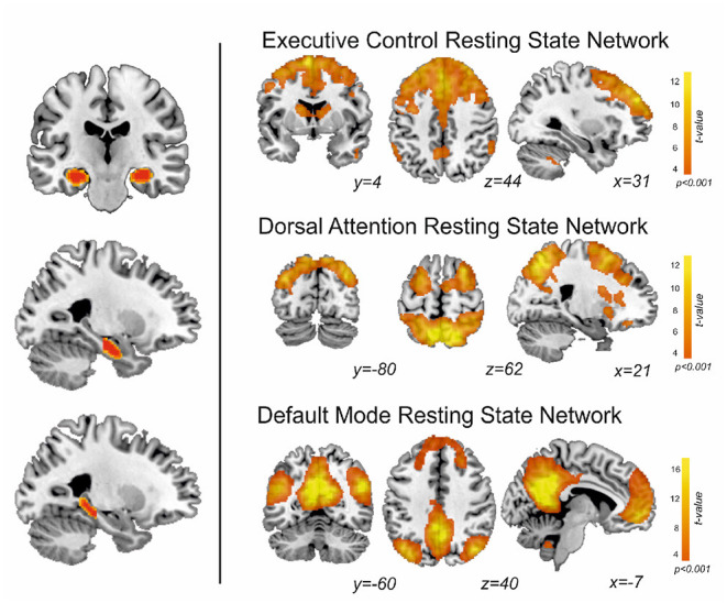
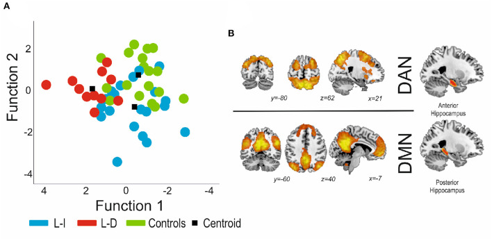
Thirty-one patients with epilepsy who underwent an anterior temporal lobe resection were pre-surgically scanned at 3-tesla, and pre/post-surgery evaluated for learning deficits using the Rey Auditory Verbal Learning Task (RAVLT). Eighteen controls matched by age, gender and handedness were also scanned and evaluated with the RAVLT. We studied the functional connectivity along the (anterior/posterior) long axis hippocampal subregions and resting-state functionally-defined brain networks involved in learning [executive (EXE), dorsal attention (DAN) and default-mode (DMN) networks]. Functional connectivity differences between the two groups of patients (learning intact or with learning decline) and controls were investigated with MANOVA and discriminant analysis.
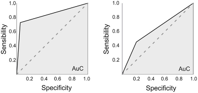
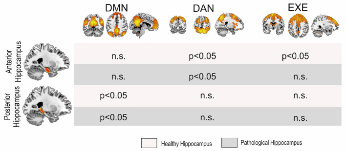
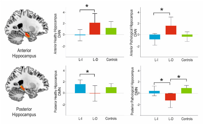
There were significant differences in the pattern of hippocampal connectivity among the groups. Regarding the anterior connectivity hippocampal pattern, our data showed an increase of connectivity in the pathological side with the DAN ( = 0.011) and the EXE ( = 0.008) when comparing learning-decline vs. learning-intact patients. Moreover, the non-pathological side showed an increase in the anterior connectivity pattern with the DAN ( = 0.027) between learning-decline vs. learning-intact patients. In contrast, the posterior hippocampus showed a reduction of connectivity in the learning-decline patients with the DMN, both in the pathological ( = 0.004) and the non-pathological sides ( = 0.036). Finally, the discriminant analysis based on the pre-operative connectivity pattern significantly differentiated the learning-decline patients from the other groups ( = 0.019).
Our findings reveal bilateral connectivity disruptions along the longitudinal axis of the hippocampi with resting-state networks, which could be key to identify those patients at risk of verbal learning decline after epilepsy surgery.
内侧颞叶切除术后20%-40%的患者学习新言语信息的能力可能受损。近年来,将癫痫理解为一种脑网络疾病,并研究大规模静息网络与认知之间的关系取得了一些进展。相关研究表明,海马与这些大规模网络的连接完整性与认知相关,有证据显示双侧海马长轴上存在功能和结构异质性。
我们的目的是研究术前沿海马长轴的静息态连接是否与颞叶前切除术后言语学习能力下降有关。
31例接受颞叶前切除术的癫痫患者在术前进行3特斯拉扫描,并在手术前后使用雷伊听觉言语学习测验(RAVLT)评估学习缺陷。还对18名年龄、性别和利手相匹配的对照者进行扫描,并使用RAVLT进行评估。我们研究了沿海马亚区(前/后)长轴的功能连接以及参与学习的静息态功能定义脑网络[执行(EXE)、背侧注意(DAN)和默认模式(DMN)网络]。采用多变量方差分析和判别分析研究两组患者(学习能力正常或下降)与对照者之间的功能连接差异。
各组之间海马连接模式存在显著差异。关于海马前连接模式,我们的数据显示,与学习能力正常的患者相比,学习能力下降的患者病变侧与DAN(P=0.011)和EXE(P=0.008)的连接增加。此外,在学习能力下降与学习能力正常的患者之间,非病变侧与DAN的前连接模式增加(P=0.027)。相比之下,学习能力下降的患者海马后部与DMN的连接在病变侧(P=0.004)和非病变侧(P=0.036)均减少。最后,基于术前连接模式的判别分析显著区分了学习能力下降的患者与其他组(P=0.019)。
我们的研究结果揭示了海马纵轴与静息态网络的双侧连接中断,这可能是识别癫痫手术后有言语学习能力下降风险患者的关键。