• 文献检索
  • 文档翻译
  • 深度研究
  • 学术资讯
  • Suppr Zotero 插件Zotero 插件
  • 邀请有礼
  • 套餐&价格
  • 历史记录
应用&插件
Suppr Zotero 插件Zotero 插件浏览器插件Mac 客户端Windows 客户端微信小程序
定价
高级版会员购买积分包购买API积分包
服务
文献检索文档翻译深度研究API 文档MCP 服务
关于我们
关于 Suppr公司介绍联系我们用户协议隐私条款
关注我们

Suppr 超能文献

核心技术专利:CN118964589B侵权必究
粤ICP备2023148730 号-1Suppr @ 2026

文献检索

告别复杂PubMed语法,用中文像聊天一样搜索,搜遍4000万医学文献。AI智能推荐,让科研检索更轻松。

立即免费搜索

文件翻译

保留排版,准确专业,支持PDF/Word/PPT等文件格式,支持 12+语言互译。

免费翻译文档

深度研究

AI帮你快速写综述,25分钟生成高质量综述,智能提取关键信息,辅助科研写作。

立即免费体验

用于附件包块分类的卵巢附件报告数据系统(O-RADS):一项系统评价和Meta分析

Ovarian Adnexal Reporting Data System (O-RADS) for Classifying Adnexal Masses: A Systematic Review and Meta-Analysis.

作者信息

Vara Julio, Manzour Nabil, Chacón Enrique, López-Picazo Ana, Linares Marta, Pascual Maria Ángela, Guerriero Stefano, Alcázar Juan Luis

机构信息

Department of Obstetrics and Gynecology, Clínica Universidad de Navarra, 31008 Pamplona, Spain.

Department of Obstetrics and Gynecology, Universitiy Hospital Puerta del Mar, 11009 Cadiz, Spain.

出版信息

Cancers (Basel). 2022 Jun 27;14(13):3151. doi: 10.3390/cancers14133151.

DOI:10.3390/cancers14133151
PMID:35804924
原文链接:https://pmc.ncbi.nlm.nih.gov/articles/PMC9264796/
Abstract

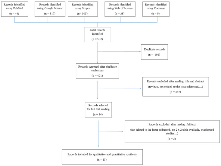
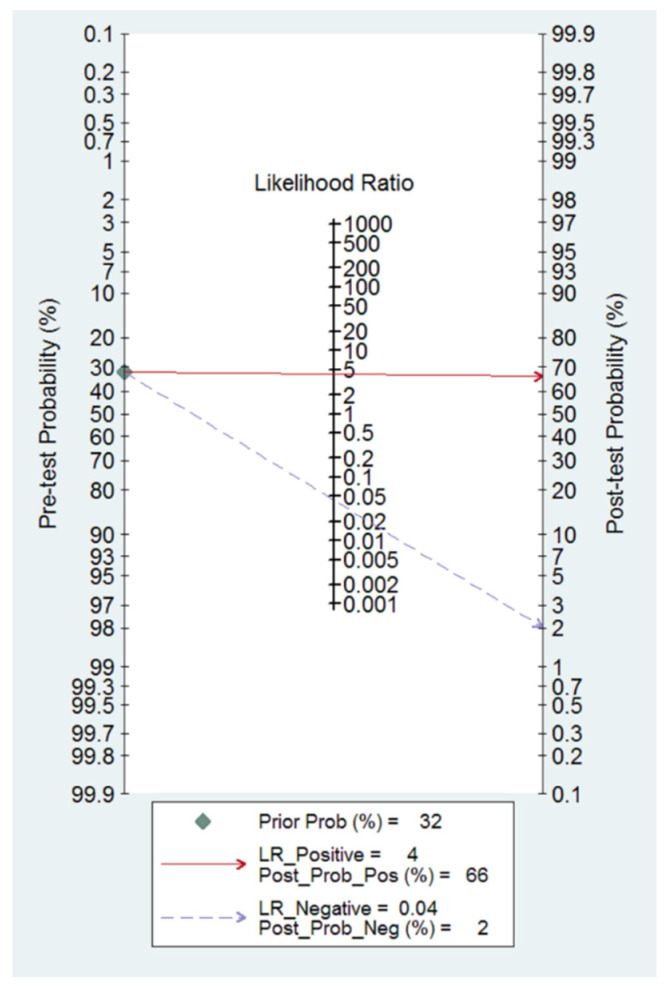
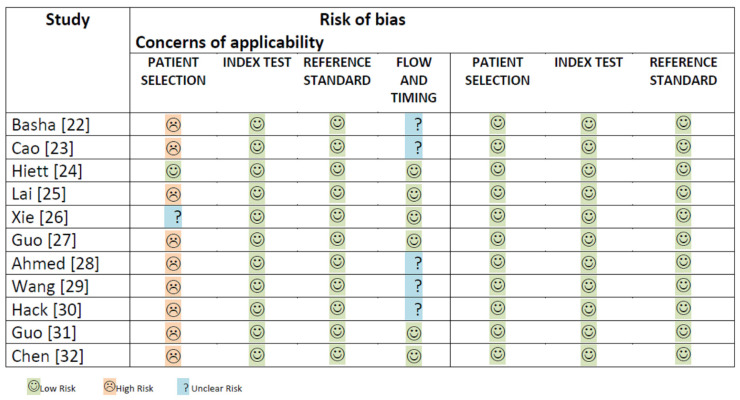
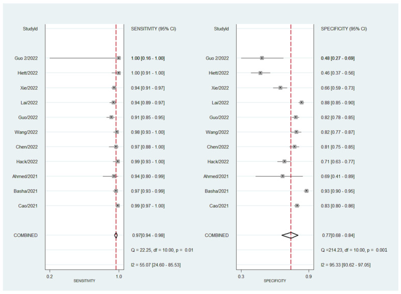
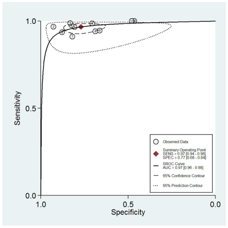
In this systematic review and meta-analysis, we aimed to assess the pooled diagnostic performance of the so-called Ovarian Adnexal Report Data System (O-RADS) for classifying adnexal masses using transvaginal ultrasound, a classification system that was introduced in 2020. We performed a search for studies reporting the use of the O-RADS system for classifying adnexal masses from January 2020 to April 2022 in several databases (Medline (PubMed), Google Scholar, Scopus, Cochrane, and Web of Science). We selected prospective and retrospective cohort studies using the O-RADS system for classifying adnexal masses with histologic diagnosis or conservative management demonstrating spontaneous resolution or persistence in cases of benign appearing masses after follow-up scan as the reference standard. We excluded studies not related to the topic under review, studies not addressing O-RADS classification, studies addressing MRI O-RADS classification, letters to the editor, commentaries, narrative reviews, consensus documents, and studies where data were not available for constructing a 2 × 2 table. The pooled sensitivity, specificity, positive and negative likelihood ratios, and diagnostic odds ratio (DOR) were calculated. The quality of the studies was evaluated using QUADAS-2. A total of 502 citations were identified. Ultimately, 11 studies comprising 4634 masses were included. The mean prevalence of ovarian malignancy was 32%. The risk of bias was high in eight studies for the "patient selection" domain. The risk of bias was low for the "index test" and "reference test" domains for all studies. Overall, the pooled estimated sensitivity, specificity, positive likelihood ratio, negative likelihood ratio, and DOR of the O-RADS system for classifying adnexal masses were 97% (95% confidence interval (CI) = 94%-98%), 77% (95% CI = 68%-84%), 4.2 (95% CI = 2.9-6.0), 0.04 (95% CI = 0.03-0.07), and 96 (95% CI = 50-185), respectively. Heterogeneity was moderate for sensitivity and high for specificity. In conclusion, the O-RADS system has good sensitivity and moderate specificity for classifying adnexal masses.

摘要

在这项系统评价和荟萃分析中,我们旨在评估所谓的卵巢附件报告数据系统(O-RADS)在使用经阴道超声对附件包块进行分类时的综合诊断性能,该分类系统于2020年推出。我们在多个数据库(Medline(PubMed)、谷歌学术、Scopus、Cochrane和科学网)中检索了2020年1月至2022年4月期间报告使用O-RADS系统对附件包块进行分类的研究。我们选择前瞻性和回顾性队列研究,这些研究使用O-RADS系统对附件包块进行分类,并以组织学诊断或保守治疗作为参考标准,对于表现为良性的包块,通过随访扫描显示自发消退或持续存在。我们排除了与所审查主题无关的研究、未涉及O-RADS分类的研究、涉及MRI O-RADS分类的研究、给编辑的信、评论、叙述性综述、共识文件以及无法获取数据以构建2×2表格的研究。计算了合并敏感性、特异性、阳性和阴性似然比以及诊断比值比(DOR)。使用QUADAS-2评估研究质量。共识别出502篇引文。最终,纳入了11项研究,共4634个包块。卵巢恶性肿瘤的平均患病率为32%。八项研究在“患者选择”领域的偏倚风险较高。所有研究在“索引测试”和“参考测试”领域的偏倚风险较低。总体而言,O-RADS系统对附件包块进行分类的合并估计敏感性、特异性、阳性似然比、阴性似然比和DOR分别为97%(95%置信区间(CI)=94%-98%)、77%(95%CI=68%-84%)、4.2(95%CI=2.9-6.0)、0.04(95%CI=0.03-0.07)和96(95%CI=50-185)。敏感性的异质性为中等,特异性的异质性较高。总之,O-RADS系统在对附件包块进行分类时具有良好的敏感性和中等的特异性。

https://cdn.ncbi.nlm.nih.gov/pmc/blobs/2908/9264796/b725afb935b3/cancers-14-03151-g005.jpg
https://cdn.ncbi.nlm.nih.gov/pmc/blobs/2908/9264796/030667419dbe/cancers-14-03151-g001.jpg
https://cdn.ncbi.nlm.nih.gov/pmc/blobs/2908/9264796/7de65566ad4f/cancers-14-03151-g002.jpg
https://cdn.ncbi.nlm.nih.gov/pmc/blobs/2908/9264796/ab724acb17f1/cancers-14-03151-g003.jpg
https://cdn.ncbi.nlm.nih.gov/pmc/blobs/2908/9264796/95313ec85a94/cancers-14-03151-g004.jpg
https://cdn.ncbi.nlm.nih.gov/pmc/blobs/2908/9264796/b725afb935b3/cancers-14-03151-g005.jpg

相似文献

1
Ovarian Adnexal Reporting Data System (O-RADS) for Classifying Adnexal Masses: A Systematic Review and Meta-Analysis.用于附件包块分类的卵巢附件报告数据系统(O-RADS):一项系统评价和Meta分析
Cancers (Basel). 2022 Jun 27;14(13):3151. doi: 10.3390/cancers14133151.
2
Gynecologic Imaging and Reporting Data System for classifying adnexal masses.妇科影像学及报告数据系统用于附件包块分类。
Minerva Obstet Gynecol. 2023 Feb;75(1):69-79. doi: 10.23736/S2724-606X.22.05122-3.
3
GI-RADS versus O-RADS in the differential diagnosis of adnexal masses: a systematic review and head-to-head meta-analysis.GI-RADS与O-RADS在附件包块鉴别诊断中的应用:一项系统评价与直接比较的荟萃分析
Ultrasonography. 2024 Nov;43(6):438-447. doi: 10.14366/usg.24105. Epub 2024 Sep 2.
4
Systematic Review and Meta-Analysis of O-RADS Ultrasound and O-RADS MRI for Risk Assessment of Ovarian and Adnexal Lesions.O-RADS 超声和 O-RADS MRI 用于卵巢和附件病变风险评估的系统评价和荟萃分析。
AJR Am J Roentgenol. 2023 Jul;221(1):21-33. doi: 10.2214/AJR.22.28396. Epub 2023 Feb 1.
5
Diagnostic performance of contrast-enhanced ultrasound (CEUS) combined with Ovarian-Adnexal Reporting and Data System (O-RADS) ultrasound risk stratification for adnexal masses: a systematic review and meta-analysis.超声造影(CEUS)联合卵巢附件报告和数据系统(O-RADS)超声风险分层对附件包块的诊断性能:一项系统评价和荟萃分析
Clin Radiol. 2024 Sep;79(9):e1167-e1175. doi: 10.1016/j.crad.2024.05.021. Epub 2024 Jun 7.
6
Diagnostic performance of the O-RADS MRI system for magnetic resonance imaging in discriminating benign and malignant adnexal lesions: a systematic review, meta-analysis, and meta-regression.O-RADS MRI系统在鉴别附件区良恶性病变的磁共振成像中的诊断性能:一项系统评价、荟萃分析和荟萃回归分析
Diagn Interv Radiol. 2025 Apr 28;31(3):171-179. doi: 10.4274/dir.2024.242784. Epub 2024 Jul 8.
7
The diagnostic performance of the Gynecologic Imaging Reporting and Data System (GI-RADS) in adnexal masses.妇科影像报告和数据系统(GI-RADS)对附件包块的诊断效能
Ann Transl Med. 2021 Mar;9(5):398. doi: 10.21037/atm-20-5170.
8
GI-RADS reporting system for ultrasound evaluation of adnexal masses in clinical practice: a prospective multicenter study.妇科附件包块超声评估的 GI-RADS 报告系统:一项前瞻性多中心研究。
Ultrasound Obstet Gynecol. 2011 Oct;38(4):450-5. doi: 10.1002/uog.9012.
9
Evaluation of American College of Radiology Ovarian-Adnexal Reporting and Data System ultrasound to predict malignancy risk in adnexal lesions.评价美国放射学会卵巢-附件报告和数据系统超声在附件病变恶性风险预测中的作用。
J Obstet Gynaecol Res. 2024 Feb;50(2):225-232. doi: 10.1111/jog.15831. Epub 2023 Nov 21.
10
External Validation of the Ovarian-Adnexal Reporting and Data System (O-RADS) Lexicon and the International Ovarian Tumor Analysis 2-Step Strategy to Stratify Ovarian Tumors Into O-RADS Risk Groups.卵巢-附件报告和数据系统 (O-RADS) 词汇表以及国际卵巢肿瘤分析两步策略的外部验证,以将卵巢肿瘤分层为 O-RADS 风险组。
JAMA Oncol. 2023 Feb 1;9(2):225-233. doi: 10.1001/jamaoncol.2022.5969.

引用本文的文献

1
Navigating Ovarian-Adnexal Reporting and Data System Magnetic Resonance Imaging (O-RADS MRI): A Review of Its Evolution, Current Advances, and Persistent Challenges in Ovarian Imaging.解读卵巢附件报告和数据系统磁共振成像(O-RADS MRI):卵巢成像中其演变、当前进展及持续挑战的综述
Cureus. 2025 Jun 25;17(6):e86717. doi: 10.7759/cureus.86717. eCollection 2025 Jun.
2
Accurate prediction of benign and malignant adnexal tumors in surgical resection and conservative treatment: construction and external validation of a diagnostic model based on CEUS, HE4, and O-RADS US v2022 evaluation.手术切除和保守治疗中附件良恶性肿瘤的准确预测:基于超声造影、人附睾蛋白4(HE4)和O-RADS US v2022评估的诊断模型构建及外部验证
J Ovarian Res. 2025 Jun 6;18(1):123. doi: 10.1186/s13048-025-01707-1.
3

本文引用的文献

1
Comparison of the O-RADS and ADNEX models regarding malignancy rate and validity in evaluating adnexal lesions.比较 O-RADS 和 ADNEX 模型在评估附件病变中的恶性率和有效性。
Eur Radiol. 2022 Nov;32(11):7854-7864. doi: 10.1007/s00330-022-08803-6. Epub 2022 May 18.
2
External Validation of O-RADS US Risk Stratification and Management System.O-RADS US 风险分层和管理系统的外部验证。
Radiology. 2022 Jul;304(1):114-120. doi: 10.1148/radiol.211868. Epub 2022 Apr 19.
3
Value of Assessment of Different Neoplasias in the Adnexa in the Differential Diagnosis of Malignant Ovarian Tumor and Benign Ovarian Tumor: A Meta-analysis.
A nomogram combining clinical features, O-RADS US, and radiomics based on ultrasound imaging for diagnosing ovarian cancer.一种基于超声成像,结合临床特征、O-RADS US和影像组学的列线图,用于诊断卵巢癌。
Sci Rep. 2025 Jun 2;15(1):19279. doi: 10.1038/s41598-025-02776-4.
4
Can transabdominal shear wave elastography play a role in solving the dilemma of complex cystic and solid ovarian tumors by ultrasound?经腹剪切波弹性成像在解决超声诊断复杂囊性及实性卵巢肿瘤的困境中能发挥作用吗?
J Ultrasound. 2025 May 31. doi: 10.1007/s40477-025-01027-6.
5
Efficacy of a combination of O-RADS, CEUS, and CA125, in identification of ovary-adnexal malignant lesions.O-RADS、超声造影(CEUS)和CA125联合应用在识别卵巢附件恶性病变中的效能
Am J Cancer Res. 2025 Feb 15;15(2):631-642. doi: 10.62347/JHJJ1149. eCollection 2025.
6
Automatic segmentation model and machine learning model grounded in ultrasound radiomics for distinguishing between low malignant risk and intermediate-high malignant risk of adnexal masses.基于超声影像组学的自动分割模型和机器学习模型,用于区分附件包块的低恶性风险和中高恶性风险。
Insights Imaging. 2025 Jan 13;16(1):14. doi: 10.1186/s13244-024-01874-7.
7
Updates on Imaging of Common Urogenital Neoplasms.常见泌尿生殖系统肿瘤的影像学进展
Cancers (Basel). 2024 Dec 30;17(1):84. doi: 10.3390/cancers17010084.
8
Ovarian-Adnexal Imaging-Reporting and Data System (O-RADS) ultrasound version 2019: a prospective validation and comparison to updated version (v2022) in pathologically confirmed adnexal masses.2019年版卵巢附件影像报告和数据系统(O-RADS)超声:对经病理证实的附件肿块进行前瞻性验证并与更新版本(2022版)进行比较
Eur Radiol. 2025 Jun;35(6):3080-3095. doi: 10.1007/s00330-024-11235-z. Epub 2024 Nov 28.
9
GI-RADS versus O-RADS in the differential diagnosis of adnexal masses: a systematic review and head-to-head meta-analysis.GI-RADS与O-RADS在附件包块鉴别诊断中的应用:一项系统评价与直接比较的荟萃分析
Ultrasonography. 2024 Nov;43(6):438-447. doi: 10.14366/usg.24105. Epub 2024 Sep 2.
10
Clinical value of ACR O-RADS combined with CA125 in the risk stratification of adnexal masses.ACR O-RADS联合CA125在附件包块风险分层中的临床价值。
Front Oncol. 2024 Aug 30;14:1369900. doi: 10.3389/fonc.2024.1369900. eCollection 2024.
附件中不同肿瘤评估在良恶性卵巢肿瘤鉴别诊断中的价值:Meta 分析。
Ultrasound Med Biol. 2022 May;48(5):730-742. doi: 10.1016/j.ultrasmedbio.2022.02.001. Epub 2022 Mar 7.
4
A comparison of the diagnostic performance of the O-RADS, RMI4, IOTA LR2, and IOTA SR systems by senior and junior doctors.资深医生和初级医生对O-RADS、RMI4、IOTA LR2和IOTA SR系统诊断性能的比较。
Ultrasonography. 2022 Jul;41(3):511-518. doi: 10.14366/usg.21237. Epub 2022 Jan 31.
5
Efficacy of IOTA simple rules, O-RADS, and CA125 to distinguish benign and malignant adnexal masses.IOTA 简单规则、O-RADS 和 CA125 对鉴别附件区良恶性肿块的效能。
J Ovarian Res. 2022 Jan 23;15(1):15. doi: 10.1186/s13048-022-00947-9.
6
Ultrasound Findings and O-RADS Malignancy Risk Stratification of Ovarian Collision Tumors.卵巢碰撞肿瘤的超声表现和 O-RADS 恶性风险分层。
J Ultrasound Med. 2022 Sep;41(9):2325-2331. doi: 10.1002/jum.15919. Epub 2021 Dec 21.
7
Comparison of O-RADS, GI-RADS, and ADNEX for Diagnosis of Adnexal Masses: An External Validation Study Conducted by Junior Sonologists.O-RADS、GI-RADS和ADNEX在附件肿物诊断中的比较:初级超声科医生进行的外部验证研究
J Ultrasound Med. 2022 Jun;41(6):1497-1507. doi: 10.1002/jum.15834. Epub 2021 Sep 21.
8
Performance of IOTA Simple Rules, Simple Rules risk assessment, ADNEX model and O-RADS in differentiating between benign and malignant adnexal lesions in North American women.IOTA简易规则、简易规则风险评估、ADNEX模型和O-RADS在北美女性附件区良恶性病变鉴别中的表现
Ultrasound Obstet Gynecol. 2022 May;59(5):668-676. doi: 10.1002/uog.24777. Epub 2022 Apr 8.
9
Diagnostic Accuracy of the ADNEX Model for Ovarian Cancer at the 15% Cut-Off Value: A Systematic Review and Meta-Analysis.ADNEX模型在15%截断值时对卵巢癌的诊断准确性:一项系统评价和Meta分析
Front Oncol. 2021 Jun 17;11:684257. doi: 10.3389/fonc.2021.684257. eCollection 2021.
10
ESGO/ISUOG/IOTA/ESGE Consensus Statement on pre-operative diagnosis of ovarian tumors.欧洲妇科肿瘤学会(ESGO)/国际妇产科超声学会(ISUOG)/国际妇产科联盟肿瘤学组(IOTA)/欧洲妇科内镜学会(ESGE)关于卵巢肿瘤术前诊断的共识声明。
Int J Gynecol Cancer. 2021 Jul;31(7):961-982. doi: 10.1136/ijgc-2021-002565. Epub 2021 Jun 10.