• 文献检索
  • 文档翻译
  • 深度研究
  • 学术资讯
  • Suppr Zotero 插件Zotero 插件
  • 邀请有礼
  • 套餐&价格
  • 历史记录
应用&插件
Suppr Zotero 插件Zotero 插件浏览器插件Mac 客户端Windows 客户端微信小程序
定价
高级版会员购买积分包购买API积分包
服务
文献检索文档翻译深度研究API 文档MCP 服务
关于我们
关于 Suppr公司介绍联系我们用户协议隐私条款
关注我们

Suppr 超能文献

核心技术专利:CN118964589B侵权必究
粤ICP备2023148730 号-1Suppr @ 2026

文献检索

告别复杂PubMed语法,用中文像聊天一样搜索,搜遍4000万医学文献。AI智能推荐,让科研检索更轻松。

立即免费搜索

文件翻译

保留排版,准确专业,支持PDF/Word/PPT等文件格式,支持 12+语言互译。

免费翻译文档

深度研究

AI帮你快速写综述,25分钟生成高质量综述,智能提取关键信息,辅助科研写作。

立即免费体验

资深医生和初级医生对O-RADS、RMI4、IOTA LR2和IOTA SR系统诊断性能的比较。

A comparison of the diagnostic performance of the O-RADS, RMI4, IOTA LR2, and IOTA SR systems by senior and junior doctors.

作者信息

Guo Yuyang, Zhao Baihua, Zhou Shan, Wen Lieming, Liu Jieyu, Fu Yaqian, Xu Fang, Liu Minghui

机构信息

Department of Ultrasound Diagnosis, The Second Xiangya Hospital, Central South University, Changsha, China.

Health Management Center, The Second Xiangya Hospital, Central South University, Changsha, China.

出版信息

Ultrasonography. 2022 Jul;41(3):511-518. doi: 10.14366/usg.21237. Epub 2022 Jan 31.

DOI:10.14366/usg.21237
PMID:35196832
原文链接:https://pmc.ncbi.nlm.nih.gov/articles/PMC9262660/
Abstract

PURPOSE

This study compared the diagnostic performance of the Ovarian-Adnexal Reporting and Data System (O-RADS), the Risk of Malignancy Index 4 (RMI4), the International Ovarian of Tumor Analysis Logistic Regression Model 2 (IOTA LR2), and the IOTA Simple Rules (IOTA SR) in predicting the malignancy of adnexal masses (AMs).

METHODS

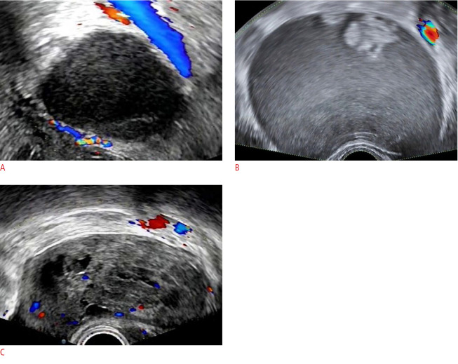
This retrospective study included 575 women with AMs between 2017 and 2020. All clinical messages, ultrasound images, and pathological findings were collected. Two senior doctors (group I) and two junior doctors (group II) used the four systems to classify AMs. The postoperative pathological diagnosis was used as the gold standard to evaluate the diagnostic efficiency. A receiver operating characteristic curve was used to test the diagnostic performance. The interrater agreement between the two groups was tested using kappa values.

RESULTS

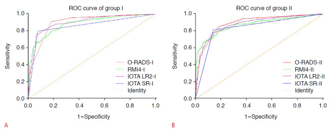
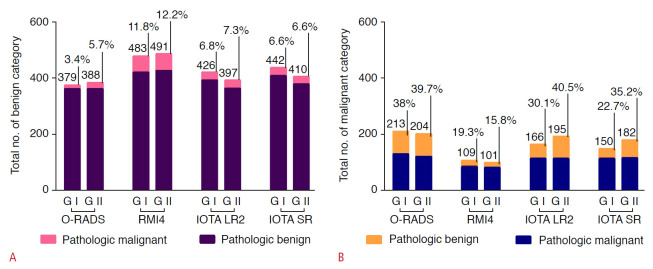
Of all 592 AMs, 447 (75.5%) were benign, 123 (20.8%) were malignant, and 22 (3.7%) were borderline. The intergroup consistency test yielded kappa values of 0.71, 0.92, 0.68, and 0.77 for the O-RADS, RMI4, IOTA LR2, and IOTA SR, respectively. To predict malignant lesions, the areas under the curve of the O-RADS, RMI4, IOTA LR2, and IOTA SR systems were 0.90, 0.89, 0.90, and 0.86 for group I and 0.89, 0.87, 0.88, and 0.84 for group II, respectively. The O-RADS had the highest sensitivity (91.0% in group I and 84.8% in group II).

CONCLUSION

The four diagnostic systems could compensate for junior doctors' inexperience in predicting malignant adnexal lesions. The O-RADS performed best and showed the highest sensitivity.

摘要

目的

本研究比较了卵巢附件报告和数据系统(O-RADS)、恶性风险指数4(RMI4)、国际卵巢肿瘤分析逻辑回归模型2(IOTA LR2)和IOTA简单规则(IOTA SR)在预测附件包块(AM)恶性程度方面的诊断性能。

方法

这项回顾性研究纳入了2017年至2020年间575例患有AM的女性。收集了所有临床信息、超声图像和病理结果。两名资深医生(第一组)和两名初级医生(第二组)使用这四种系统对AM进行分类。术后病理诊断作为评估诊断效率的金标准。采用受试者工作特征曲线来测试诊断性能。使用kappa值来测试两组之间的评分者间一致性。

结果

在所有592个AM中,447个(75.5%)为良性,123个(20.8%)为恶性,22个(3.7%)为交界性。O-RADS、RMI4、IOTA LR2和IOTA SR的组间一致性测试kappa值分别为0.71、0.92、0.68和0.77。为预测恶性病变,O-RADS、RMI4、IOTA LR2和IOTA SR系统在第一组中的曲线下面积分别为0.90、0.89、0.90和0.86,在第二组中分别为0.89、0.87、0.88和0.84。O-RADS具有最高的敏感性(第一组为91.0%,第二组为84.8%)。

结论

这四种诊断系统可以弥补初级医生在预测附件恶性病变方面的经验不足。O-RADS表现最佳,敏感性最高。

https://cdn.ncbi.nlm.nih.gov/pmc/blobs/b17f/9262660/016a189db602/usg-21237f3.jpg
https://cdn.ncbi.nlm.nih.gov/pmc/blobs/b17f/9262660/5b9c09f7d344/usg-21237f1.jpg
https://cdn.ncbi.nlm.nih.gov/pmc/blobs/b17f/9262660/668ad2c6ebd9/usg-21237f2.jpg
https://cdn.ncbi.nlm.nih.gov/pmc/blobs/b17f/9262660/016a189db602/usg-21237f3.jpg
https://cdn.ncbi.nlm.nih.gov/pmc/blobs/b17f/9262660/5b9c09f7d344/usg-21237f1.jpg
https://cdn.ncbi.nlm.nih.gov/pmc/blobs/b17f/9262660/668ad2c6ebd9/usg-21237f2.jpg
https://cdn.ncbi.nlm.nih.gov/pmc/blobs/b17f/9262660/016a189db602/usg-21237f3.jpg

相似文献

1
A comparison of the diagnostic performance of the O-RADS, RMI4, IOTA LR2, and IOTA SR systems by senior and junior doctors.资深医生和初级医生对O-RADS、RMI4、IOTA LR2和IOTA SR系统诊断性能的比较。
Ultrasonography. 2022 Jul;41(3):511-518. doi: 10.14366/usg.21237. Epub 2022 Jan 31.
2
Comparison of diagnostic efficiency between IOTA LR2 model and doctors experiences.IOTA LR2 模型与医生经验的诊断效率比较。
Zhong Nan Da Xue Xue Bao Yi Xue Ban. 2022 Aug 28;47(8):1082-1088. doi: 10.11817/j.issn.1672-7347.2022.220051.
3
Performance of IOTA Simple Rules, Simple Rules risk assessment, ADNEX model and O-RADS in differentiating between benign and malignant adnexal lesions in North American women.IOTA简易规则、简易规则风险评估、ADNEX模型和O-RADS在北美女性附件区良恶性病变鉴别中的表现
Ultrasound Obstet Gynecol. 2022 May;59(5):668-676. doi: 10.1002/uog.24777. Epub 2022 Apr 8.
4
Efficacy of IOTA simple rules, O-RADS, and CA125 to distinguish benign and malignant adnexal masses.IOTA 简单规则、O-RADS 和 CA125 对鉴别附件区良恶性肿块的效能。
J Ovarian Res. 2022 Jan 23;15(1):15. doi: 10.1186/s13048-022-00947-9.
5
Diagnostic performance of IOTA SR and O-RADS combined with CA125, HE4, and risk of malignancy algorithm to distinguish benign and malignant adnexal masses.IOTA SR 和 O-RADS 联合 CA125、HE4 和恶性风险算法对附件肿块良恶性的诊断性能。
Eur J Radiol. 2023 Aug;165:110926. doi: 10.1016/j.ejrad.2023.110926. Epub 2023 Jun 16.
6
Comparative diagnostic accuracy of the IOTA SRR and LR2 scoring systems for discriminating between malignant and Benign Adnexal masses by junior physicians in Chinese patients: a retrospective observational study.中文医师应用 IOTA SRR 和 LR2 评分系统鉴别中国患者良恶性附件包块的对比诊断准确性:一项回顾性观察性研究。
BMC Womens Health. 2023 Nov 8;23(1):585. doi: 10.1186/s12905-023-02719-z.
7
Comparison of the diagnostic efficiency between the O-RADS US risk stratification system and doctors' subjective judgment.O-RADS US 风险分层系统与医生主观判断的诊断效能比较。
BMC Med Imaging. 2023 Nov 20;23(1):190. doi: 10.1186/s12880-023-01153-9.
8
Differentiating Benign From Malignant Ovarian Masses With Solid Components: Diagnostic Performance of CEUS Combined With IOTA Simple Rules and O-RADS.超声造影联合 IOTA 简单规则和 O-RADS 对卵巢实性成分良恶性肿块的鉴别诊断价值
Ultrasound Med Biol. 2024 Sep;50(9):1449-1458. doi: 10.1016/j.ultrasmedbio.2024.05.024. Epub 2024 Jun 13.
9
Lesion size affects diagnostic performance of IOTA logistic regression models, IOTA simple rules and risk of malignancy index in discriminating between benign and malignant adnexal masses.病灶大小影响 IOTA 逻辑回归模型、IOTA 简单规则和恶性风险指数在鉴别附件包块良恶性方面的诊断性能。
Ultrasound Obstet Gynecol. 2012 Sep;40(3):345-54. doi: 10.1002/uog.11167. Epub 2012 Aug 7.
10
Comparison of O-RADS, GI-RADS, and IOTA simple rules regarding malignancy rate, validity, and reliability for diagnosis of adnexal masses.附件包块良恶性诊断中 O-RADS、GI-RADS 和 IOTA 简单规则的比较:恶性率、有效性和可靠性。
Eur Radiol. 2021 Feb;31(2):674-684. doi: 10.1007/s00330-020-07143-7. Epub 2020 Aug 18.

引用本文的文献

1
Development and Validation of Differential Diagnosis Models and Nomograms Based on Serum D-Dimer and Other Multimodal Information for Borderline and Benign Epithelial Ovarian Tumors: A Multicenter Study.基于血清D-二聚体及其他多模态信息的交界性和良性上皮性卵巢肿瘤鉴别诊断模型及列线图的开发与验证:一项多中心研究
Diagnostics (Basel). 2025 Aug 14;15(16):2035. doi: 10.3390/diagnostics15162035.
2
O-RADS US versus IOTA simple rules in the diagnosis of benign and malignant adnexal masses: a prospective study.O-RADS超声与IOTA简单规则在附件肿块良恶性诊断中的应用:一项前瞻性研究
BMC Med Imaging. 2025 Jul 28;25(1):297. doi: 10.1186/s12880-025-01845-4.
3

本文引用的文献

1
Comparison of O-RADS, GI-RADS, and ADNEX for Diagnosis of Adnexal Masses: An External Validation Study Conducted by Junior Sonologists.O-RADS、GI-RADS和ADNEX在附件肿物诊断中的比较:初级超声科医生进行的外部验证研究
J Ultrasound Med. 2022 Jun;41(6):1497-1507. doi: 10.1002/jum.15834. Epub 2021 Sep 21.
2
Performance of IOTA Simple Rules, Simple Rules risk assessment, ADNEX model and O-RADS in differentiating between benign and malignant adnexal lesions in North American women.IOTA简易规则、简易规则风险评估、ADNEX模型和O-RADS在北美女性附件区良恶性病变鉴别中的表现
Ultrasound Obstet Gynecol. 2022 May;59(5):668-676. doi: 10.1002/uog.24777. Epub 2022 Apr 8.
3
Advancements in artificial intelligence for ultrasound diagnosis of ovarian cancer: a comprehensive review.
人工智能在卵巢癌超声诊断中的进展:一项全面综述。
Front Oncol. 2025 Jun 12;15:1581157. doi: 10.3389/fonc.2025.1581157. eCollection 2025.
4
Accurate prediction of benign and malignant adnexal tumors in surgical resection and conservative treatment: construction and external validation of a diagnostic model based on CEUS, HE4, and O-RADS US v2022 evaluation.手术切除和保守治疗中附件良恶性肿瘤的准确预测:基于超声造影、人附睾蛋白4(HE4)和O-RADS US v2022评估的诊断模型构建及外部验证
J Ovarian Res. 2025 Jun 6;18(1):123. doi: 10.1186/s13048-025-01707-1.
5
Comparison of the Diagnostic Performance of Ovarian Adnexal Reporting Data System (O-RADS) With IOTA Simple Rules and ADNEX Model for Classifying Adnexal Masses: A Head-To-Head Meta-Analysis.卵巢附件报告数据系统(O-RADS)与IOTA简单规则及ADNEX模型对附件包块进行分类的诊断性能比较:一项直接对比的Meta分析
J Clin Ultrasound. 2025 Apr 29. doi: 10.1002/jcu.24048.
6
The diagnostic performance of International Ovarian Tumor Analysis: Simple Rules for diagnosing ovarian tumors-a systematic review and meta-analysis.国际卵巢肿瘤分析的诊断性能:诊断卵巢肿瘤的简易规则——一项系统评价和荟萃分析
Front Oncol. 2025 Jan 20;14:1474930. doi: 10.3389/fonc.2024.1474930. eCollection 2024.
7
Inter-reader reliability of Ovarian-Adnexal Reporting and Data System US: a systematic review and meta-analysis.卵巢附件报告和数据系统超声检查的阅片者间可靠性:一项系统评价和荟萃分析
Abdom Radiol (NY). 2025 Jan 22. doi: 10.1007/s00261-025-04813-2.
8
Automatic segmentation model and machine learning model grounded in ultrasound radiomics for distinguishing between low malignant risk and intermediate-high malignant risk of adnexal masses.基于超声影像组学的自动分割模型和机器学习模型,用于区分附件包块的低恶性风险和中高恶性风险。
Insights Imaging. 2025 Jan 13;16(1):14. doi: 10.1186/s13244-024-01874-7.
9
Diagnostic performances of the Ovarian Adnexal Reporting and Data System, the Risk of Ovarian Malignancy Algorithm, and the Copenhagen Index in the preoperative prediction of ovarian cancer: a prospective cohort study.卵巢附件报告和数据系统、卵巢恶性肿瘤风险算法及哥本哈根指数在卵巢癌术前预测中的诊断性能:一项前瞻性队列研究。
J Gynecol Oncol. 2025 Mar;36(2):e30. doi: 10.3802/jgo.2025.36.e30. Epub 2024 Sep 24.
10
Clinical value of ACR O-RADS combined with CA125 in the risk stratification of adnexal masses.ACR O-RADS联合CA125在附件包块风险分层中的临床价值。
Front Oncol. 2024 Aug 30;14:1369900. doi: 10.3389/fonc.2024.1369900. eCollection 2024.
Diagnostic accuracy and inter-observer reliability of the O-RADS scoring system among staff radiologists in a North American academic clinical setting.
北美学术临床环境中工作人员放射科医生的 O-RADS 评分系统的诊断准确性和观察者间可靠性。
Abdom Radiol (NY). 2021 Oct;46(10):4967-4973. doi: 10.1007/s00261-021-03193-7. Epub 2021 Jun 29.
4
Comparison of the Diagnostic Performances of Ultrasound-Based Models for Predicting Malignancy in Patients With Adnexal Masses.基于超声的附件包块患者恶性肿瘤预测模型诊断性能的比较
Front Oncol. 2021 Jun 1;11:673722. doi: 10.3389/fonc.2021.673722. eCollection 2021.
5
Performance of O-RADS MRI Score for Classifying Indeterminate Adnexal Masses at US.O-RADS MRI评分在超声检查中对附件区不确定肿块进行分类的表现。
Radiol Imaging Cancer. 2021 May;3(3):e219008. doi: 10.1148/rycan.2021219008.
6
Validation of American College of Radiology Ovarian-Adnexal Reporting and Data System Ultrasound (O-RADS US): Analysis on 1054 adnexal masses.美国放射学院卵巢-附件报告和数据系统超声(O-RADS US)的验证:对 1054 个附件肿块的分析。
Gynecol Oncol. 2021 Jul;162(1):107-112. doi: 10.1016/j.ygyno.2021.04.031. Epub 2021 May 7.
7
Comparison of IOTA three-step strategy and logistic regression model LR2 for discriminating between benign and malignant adnexal masses.比较 IOTA 三步骤策略和逻辑回归模型 LR2 用于鉴别附件肿块良恶性。
Med Ultrason. 2021 May 20;23(2):168-175. doi: 10.11152/mu-2732. Epub 2020 Dec 12.
8
Comparison of O-RADS, GI-RADS, and IOTA simple rules regarding malignancy rate, validity, and reliability for diagnosis of adnexal masses.附件包块良恶性诊断中 O-RADS、GI-RADS 和 IOTA 简单规则的比较:恶性率、有效性和可靠性。
Eur Radiol. 2021 Feb;31(2):674-684. doi: 10.1007/s00330-020-07143-7. Epub 2020 Aug 18.
9
Comparison of the predictive performance of risk of malignancy indexes 1-4, HE4 and risk of malignancy algorithm in the triage of adnexal masses.比较风险恶性指数 1-4、HE4 和风险恶性算法在附件包块分诊中的预测性能。
J Ovarian Res. 2020 Apr 25;13(1):46. doi: 10.1186/s13048-020-00643-6.
10
O-RADS US Risk Stratification and Management System: A Consensus Guideline from the ACR Ovarian-Adnexal Reporting and Data System Committee.O-RADS US 风险分层与管理系统:ACR 卵巢-附件报告和数据系统委员会的共识指南。
Radiology. 2020 Jan;294(1):168-185. doi: 10.1148/radiol.2019191150. Epub 2019 Nov 5.