Department of Nephrology Affiliated Hospital of Guizhou Medical University, Guiyang, 550004 Guizhou, China.
Department of Endocrinology Affiliated Hospital of Guizhou Medical University, Guiyang, 550004 Guizhou, China.
Biomed Res Int. 2022 Jun 30;2022:9018379. doi: 10.1155/2022/9018379. eCollection 2022.
To investigate the effects of peroxisome proliferator-activated receptor (PPAR) expression on renal podocyte in diabetic mice by conditionally knockout mouse PPAR gene.
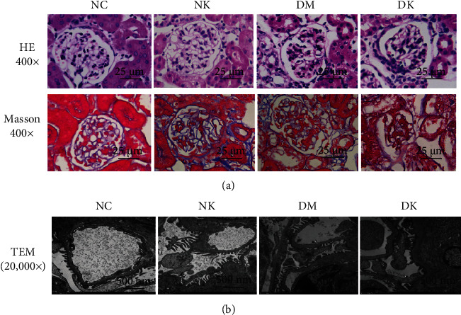
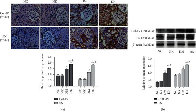
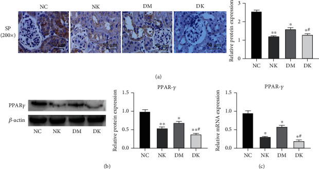
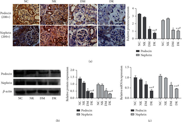
Wild-type C57BL mice and PPAR gene knockout mice were used as research objects to establish the diabetic mouse model, which was divided into normal control group (NC group), normal glucose PPAR gene knockout group (NK group), diabetic wild-type group (DM group), and diabetic PPAR gene knockout group (DK group), with 8 mice in each group. After 16 weeks, the mice were sacrificed for renal tissue collection. Morphological changes of renal tissue were observed by HE and Masson staining, and ultrastructure of renal tissue was observed by transmission electron microscope. Protein expressions of PPAR, podocin, nephrin, collagen IV, and fibronectin (FN) in renal tissues were detected by immunohistochemistry and Western blot, and mRNA changes of PPAR, podocin, and nephrin in renal tissues were detected by qRT-PCR.
Compared with the NC group, the protein and mRNA expressions of PPAR, podocin, and nephrin decreased in the kidney tissue of mice in the DM group, while the protein expressions of collagen IV and FN increased. The expression of various proteins in kidney tissues of the DK group was consistent with that of the DM group, and the difference was more obvious. The expression of PPAR protein and mRNA decreased in the NK group, while the expression of podocin, nephrin protein and mRNA, collagen IV, and FN protein showed no significant difference.
In diabetic renal tissue, the loss of PPAR can aggravate podocellular damage and thus promote the occurrence of diabetic renal fibrosis. Increasing the expression of PPAR may effectively relieve renal podocyte impairment in diabetic patients, which can be used for the treatment of diabetic nephropathy.
通过条件性敲除小鼠过氧化物酶体增殖物激活受体(PPAR)基因,研究 PPAR 表达对糖尿病小鼠肾脏足细胞的影响。
以野生型 C57BL 小鼠和 PPAR 基因敲除小鼠为研究对象,建立糖尿病小鼠模型,分为正常对照组(NC 组)、正常葡萄糖 PPAR 基因敲除组(NK 组)、糖尿病野生型组(DM 组)和糖尿病 PPAR 基因敲除组(DK 组),每组 8 只。16 周后处死小鼠,采集肾组织。HE 和 Masson 染色观察肾组织形态学变化,透射电镜观察肾组织超微结构。免疫组化和 Western blot 检测肾组织中 PPAR、足细胞裂孔膜蛋白(podocin)、nephrin、Ⅳ型胶原(collagen IV)和纤维连接蛋白(FN)的蛋白表达,qRT-PCR 检测肾组织中 PPAR、podocin 和 nephrin 的 mRNA 变化。
与 NC 组相比,DM 组小鼠肾组织中 PPAR、podocin 和 nephrin 的蛋白和 mRNA 表达降低,collagen IV 和 FN 蛋白表达增加。DK 组肾组织各蛋白表达与 DM 组一致,差异更明显。NK 组 PPAR 蛋白和 mRNA 表达降低,podocin、nephrin 蛋白和 mRNA、collagen IV、FN 蛋白表达无明显差异。
在糖尿病肾组织中,PPAR 的缺失会加重足细胞损伤,从而促进糖尿病肾纤维化的发生。增加 PPAR 的表达可能有效缓解糖尿病患者的肾足细胞损伤,可用于糖尿病肾病的治疗。