• 文献检索
  • 文档翻译
  • 深度研究
  • 学术资讯
  • Suppr Zotero 插件Zotero 插件
  • 邀请有礼
  • 套餐&价格
  • 历史记录
应用&插件
Suppr Zotero 插件Zotero 插件浏览器插件Mac 客户端Windows 客户端微信小程序
定价
高级版会员购买积分包购买API积分包
服务
文献检索文档翻译深度研究API 文档MCP 服务
关于我们
关于 Suppr公司介绍联系我们用户协议隐私条款
关注我们

Suppr 超能文献

核心技术专利:CN118964589B侵权必究
粤ICP备2023148730 号-1Suppr @ 2026

文献检索

告别复杂PubMed语法,用中文像聊天一样搜索,搜遍4000万医学文献。AI智能推荐,让科研检索更轻松。

立即免费搜索

文件翻译

保留排版,准确专业,支持PDF/Word/PPT等文件格式,支持 12+语言互译。

免费翻译文档

深度研究

AI帮你快速写综述,25分钟生成高质量综述,智能提取关键信息,辅助科研写作。

立即免费体验

脑膜瘤的影像学与组织病理学类型的相关性:一项队列研究。

Correlation between Radiological Images and Histopathological Type of Meningioma: A Cohort Study.

机构信息

Department of Neurosurgery, Faculty of Medicine, Hasanuddin University, Makassar, Indonesia.

Department of Neurosurgery, Wahidin Sudirohusodo Hospital, Makassar, Indonesia.

出版信息

Ethiop J Health Sci. 2022 May;32(3):597-604. doi: 10.4314/ejhs.v32i3.16.

DOI:10.4314/ejhs.v32i3.16
PMID:35813670
原文链接:https://pmc.ncbi.nlm.nih.gov/articles/PMC9214728/
Abstract

BACKGROUND

Histologically affirmed meningiomas represent 37.6% of all essential central nervous system tumors and half of all types of critical central nervous system tumors. This study compares computed tomography (CT) scans of the head with histological findings to establish the characteristics of different types of meningiomas observed in eastern Indonesia.

METHODS

This prospective study evaluated 224 patients by examining the correlation between histological and CT data collected from January to December 2020 at Wahidin Sudirohusodo Hospital, Makassar, Indonesia. We assessed data including the location of pre- and post-contrast CT scans, number of tumors, margin, density, contrast enhancement, bony reaction, calcification, and perifocal edema. Patients underwent biopsies followed by an examination of the anatomical pathology tissue.

RESULTS

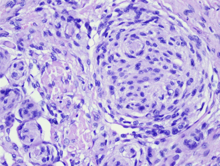
The female-to-male ratio of participants was 4.2 to 1, and the highest incidence was observed in participants of both genders aged 40-60 years. The most common meningioma subtype was meningothelial, while the most commonly observed locations involved the convexity and sphenoid regions. Most meningiomas had well-defined margins on CT imaging: 54.5% of patients exhibited isodense lesions on pre-contrast scans, and 64.7% exhibited high-contrast enhancement. Bone destruction developed in 4.1% of patients, while hyperostosis was observed in 17.4%, and calcification was present in 10.3% of the participants. Edema was identified in 65.2% of cases, of which moderate edema was the most common manifestation.

CONCLUSION

Meningioma should be highly suspected in female patients aged 40-60 with isodense lesions on pre-contrast CT scans and high-contrast enhancement on post-contrast CT scans. Meningiomas were primarily classified as convexity meningiomas with well-defined margins. The presence of hyperostosis, calcification, and brain edema supported the meningioma diagnosis.

摘要

背景

经组织学证实的脑膜瘤占所有中枢神经系统肿瘤的 37.6%,占所有中枢神经系统肿瘤的一半。本研究通过比较头部 CT 扫描与组织学发现,来确定在印度尼西亚东部观察到的不同类型脑膜瘤的特征。

方法

本前瞻性研究评估了 224 名患者,通过检查 2020 年 1 月至 12 月在印度尼西亚望加锡 Wahidin Sudirohusodo 医院收集的组织学和 CT 数据之间的相关性。我们评估了包括 CT 扫描的位置、肿瘤数量、边缘、密度、对比增强、骨反应、钙化和周围水肿等数据。患者接受了活检,随后对解剖病理组织进行了检查。

结果

参与者的男女比例为 4.2:1,发病率最高的是 40-60 岁的男女两性。最常见的脑膜瘤亚型是脑膜内皮型,最常见的观察部位是凸面和蝶骨区。大多数脑膜瘤在 CT 成像上具有清晰的边缘:54.5%的患者在增强前 CT 扫描上显示等密度病变,64.7%的患者显示高对比度增强。4.1%的患者发生骨破坏,17.4%的患者出现骨质增生,10.3%的患者出现钙化。65.2%的患者出现水肿,其中中度水肿最常见。

结论

对于增强前 CT 扫描显示等密度病变和增强后 CT 扫描显示高对比度增强的 40-60 岁女性患者,应高度怀疑脑膜瘤。脑膜瘤主要分类为边界清楚的凸面脑膜瘤。骨质增生、钙化和脑水肿的存在支持脑膜瘤的诊断。

https://cdn.ncbi.nlm.nih.gov/pmc/blobs/9816/9214728/6831c7c82e24/EJHS3203-0597Fig2.jpg
https://cdn.ncbi.nlm.nih.gov/pmc/blobs/9816/9214728/1ce355fe41a0/EJHS3203-0597Fig1.jpg
https://cdn.ncbi.nlm.nih.gov/pmc/blobs/9816/9214728/6831c7c82e24/EJHS3203-0597Fig2.jpg
https://cdn.ncbi.nlm.nih.gov/pmc/blobs/9816/9214728/1ce355fe41a0/EJHS3203-0597Fig1.jpg
https://cdn.ncbi.nlm.nih.gov/pmc/blobs/9816/9214728/6831c7c82e24/EJHS3203-0597Fig2.jpg

相似文献

1
Correlation between Radiological Images and Histopathological Type of Meningioma: A Cohort Study.脑膜瘤的影像学与组织病理学类型的相关性:一项队列研究。
Ethiop J Health Sci. 2022 May;32(3):597-604. doi: 10.4314/ejhs.v32i3.16.
2
Neuroradiological features of patients with bilateral macronodular adrenocortical disease and meningiomas associated or not with genetic variants of ARMC5- a case series.双侧巨结节性肾上腺皮质疾病伴脑膜瘤患者的神经放射学特征,伴或不伴 ARMC5 基因突变的病例系列。
J Neurooncol. 2024 Jul;168(3):405-413. doi: 10.1007/s11060-024-04680-9. Epub 2024 Apr 17.
3
Does bony hyperostosis in intracranial meningioma signify tumor invasion? A radio-pathologic study.颅内脑膜瘤的骨肥厚是否提示肿瘤侵犯?一项放射病理学研究。
Neurol India. 2012 Jan-Feb;60(1):50-4. doi: 10.4103/0028-3886.93589.
4
Usefulness of a Rim-Enhancing Pattern on the Contrast-Enhanced 3D-FLAIR Sequence and MRI Characteristics for Distinguishing Meningioma and Malignant Dural-Based Tumor.对比增强 3D-FLAIR 序列边缘增强模式和 MRI 特征在鉴别脑膜瘤和恶性硬脑膜肿瘤中的作用。
AJNR Am J Neuroradiol. 2023 Mar;44(3):247-253. doi: 10.3174/ajnr.A7780. Epub 2023 Feb 2.
5
WHO grade I meningioma subtypes: MRI features and pathological analysis.WHO 分级 I 型脑膜瘤亚型:MRI 特征与病理分析。
Life Sci. 2018 Nov 15;213:50-56. doi: 10.1016/j.lfs.2018.08.061. Epub 2018 Aug 25.
6
[X-ray computed tomographic aspects of malignant meningioma and meningeal hemangiopericytoma].[恶性脑膜瘤和脑膜血管外皮细胞瘤的X线计算机断层扫描表现]
Can Assoc Radiol J. 1996 Aug;47(4):279-87.
7
Imaging features of intracranial psammomatous meningioma.颅内砂粒体型脑膜瘤的影像学特征。
J Neuroradiol. 2017 Oct;44(6):395-399. doi: 10.1016/j.neurad.2017.06.003. Epub 2017 Jul 6.
8
Investigation of blood flow in meningothelial and fibrous meningiomas by xenon-enhanced CT scanning.通过氙增强CT扫描研究脑膜内皮型和纤维型脑膜瘤的血流情况。
Neurol Res. 2000 Sep;22(6):615-9. doi: 10.1080/01616412.2000.11740729.
9
A radiopathological classification of dural tail sign of meningiomas.脑膜瘤硬脑膜尾征的放射病理学分类。
J Neurosurg. 2012 Oct;117(4):645-53. doi: 10.3171/2012.6.JNS111987. Epub 2012 Jul 27.
10
Cranial Intraosseous Meningiomas: CT and MRI Findings.颅骨骨内脑膜瘤:CT与MRI表现
Turk Neurosurg. 2020;30(4):542-549. doi: 10.5137/1019-5149.JTN.25694-18.4.

引用本文的文献

1
Intracranial meningiomas at a tertiary hospital: Spectrum of MRI findings with histopathologic correlation.一家三级医院的颅内脑膜瘤:具有组织病理学相关性的MRI表现谱
SA J Radiol. 2024 Mar 27;28(1):2812. doi: 10.4102/sajr.v28i1.2812. eCollection 2024.
2
Atypical radiological aspect of meningioma: Web-like enhancement.脑膜瘤的非典型影像学表现:网状强化。
Radiol Case Rep. 2023 Jun 6;18(8):2796-2799. doi: 10.1016/j.radcr.2023.05.029. eCollection 2023 Aug.

本文引用的文献

1
Effects of hyperbaric oxygen therapy on the healing of thermal burns and its relationship with ICAM-1: A case-control study.高压氧疗法对热烧伤愈合的影响及其与细胞间黏附分子-1的关系:一项病例对照研究。
Ann Med Surg (Lond). 2020 Dec 25;61:104-109. doi: 10.1016/j.amsu.2020.12.025. eCollection 2021 Jan.
2
Imaging spectrum of meningiomas: a review of uncommon imaging appearances and their histopathological and prognostic significance.脑膜瘤的影像学谱:罕见影像学表现及其组织病理学和预后意义的综述
Pol J Radiol. 2019 Dec 29;84:e630-e653. doi: 10.5114/pjr.2019.92421. eCollection 2019.
3
STROCSS 2019 Guideline: Strengthening the reporting of cohort studies in surgery.
STROCSS 2019 指南:加强外科学队列研究报告。
Int J Surg. 2019 Dec;72:156-165. doi: 10.1016/j.ijsu.2019.11.002. Epub 2019 Nov 6.
4
CBTRUS Statistical Report: Primary Brain and Other Central Nervous System Tumors Diagnosed in the United States in 2012-2016.美国 2012-2016 年诊断的原发性脑和其他中枢神经系统肿瘤 CBTRUS 统计报告。
Neuro Oncol. 2019 Nov 1;21(Suppl 5):v1-v100. doi: 10.1093/neuonc/noz150.
5
Dural masses: meningiomas and their mimics.硬脑膜肿物:脑膜瘤及其类似病变
Insights Imaging. 2019 Feb 6;10(1):11. doi: 10.1186/s13244-019-0697-7.
6
Imaging and diagnostic advances for intracranial meningiomas.颅内脑膜瘤的影像学和诊断进展。
Neuro Oncol. 2019 Jan 14;21(Suppl 1):i44-i61. doi: 10.1093/neuonc/noy143.
7
An overview of meningiomas.脑膜瘤概述。
Future Oncol. 2018 Sep;14(21):2161-2177. doi: 10.2217/fon-2018-0006. Epub 2018 Aug 7.
8
CBTRUS Statistical Report: Primary Brain and Other Central Nervous System Tumors Diagnosed in the United States in 2009-2013.CBTRUS统计报告:2009 - 2013年美国原发性脑和其他中枢神经系统肿瘤诊断情况
Neuro Oncol. 2016 Oct 1;18(suppl_5):v1-v75. doi: 10.1093/neuonc/now207.
9
Meningioma recurrence.脑膜瘤复发
Open Med (Wars). 2016 Jun 9;11(1):168-173. doi: 10.1515/med-2016-0032. eCollection 2016.
10
The 2016 World Health Organization Classification of Tumors of the Central Nervous System: a summary.2016 年世界卫生组织中枢神经系统肿瘤分类:概述。
Acta Neuropathol. 2016 Jun;131(6):803-20. doi: 10.1007/s00401-016-1545-1. Epub 2016 May 9.