MRC Prion Unit at UCL, Institute of Prion Diseases, University College London, 33 Cleveland Street, London, W1W 7FF, UK.
Institute of Structural and Molecular Biology, Department of Biological Sciences, Birkbeck College, University of London, Malet Street, London, WC1E 7HX, UK.
Nat Commun. 2022 Jul 13;13(1):4004. doi: 10.1038/s41467-022-30457-7.
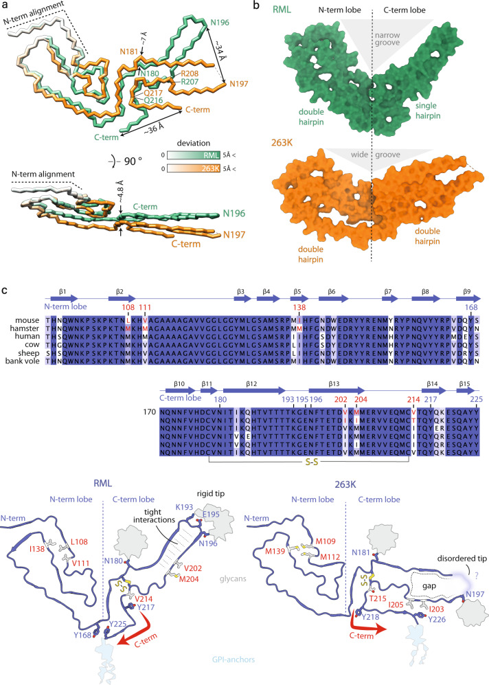
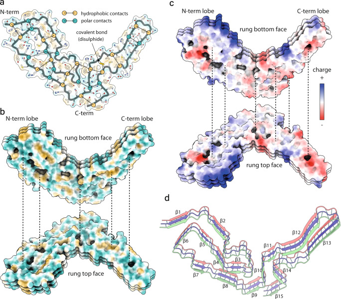
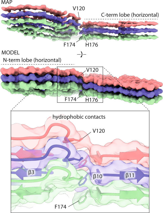
Mammalian prions propagate as distinct strains and are composed of multichain assemblies of misfolded host-encoded prion protein (PrP). Here, we present a near-atomic resolution cryo-EM structure of PrP fibrils present in highly infectious prion rod preparations isolated from the brains of RML prion-infected mice. We found that prion rods comprise single-protofilament helical amyloid fibrils that coexist with twisted pairs of the same protofilaments. Each rung of the protofilament is formed by a single PrP monomer with the ordered core comprising PrP residues 94-225, which folds to create two asymmetric lobes with the N-linked glycans and the glycosylphosphatidylinositol anchor projecting from the C-terminal lobe. The overall architecture is comparable to that of recently reported PrP fibrils isolated from the brain of hamsters infected with the 263K prion strain. However, there are marked conformational variations that could result from differences in PrP sequence and/or represent distinguishing features of the distinct prion strains.
哺乳动物朊病毒以不同的株系形式传播,由错误折叠的宿主编码朊病毒蛋白(PrP)的多链组装体组成。在这里,我们呈现了一种来自 RML 朊病毒感染小鼠大脑的高传染性朊病毒杆状制剂中存在的 PrP 原纤维的近原子分辨率冷冻电镜结构。我们发现,朊病毒杆由单原纤维螺旋状淀粉样纤维组成,这些纤维与同一原纤维的扭曲对共存。每个原纤维的梯级由单个 PrP 单体形成,有序核心包含 PrP 残基 94-225,其折叠形成两个不对称叶,N-连接的糖基和糖基磷脂酰肌醇锚定从 C 末端叶突出。整体结构与最近从感染 263K 朊病毒株的仓鼠大脑中分离出的 PrP 原纤维的报道相似。然而,存在明显的构象变化,这可能是由于 PrP 序列的差异,或者代表不同朊病毒株系的区别特征。