• 文献检索
  • 文档翻译
  • 深度研究
  • 学术资讯
  • Suppr Zotero 插件Zotero 插件
  • 邀请有礼
  • 套餐&价格
  • 历史记录
应用&插件
Suppr Zotero 插件Zotero 插件浏览器插件Mac 客户端Windows 客户端微信小程序
定价
高级版会员购买积分包购买API积分包
服务
文献检索文档翻译深度研究API 文档MCP 服务
关于我们
关于 Suppr公司介绍联系我们用户协议隐私条款
关注我们

Suppr 超能文献

核心技术专利:CN118964589B侵权必究
粤ICP备2023148730 号-1Suppr @ 2026

文献检索

告别复杂PubMed语法,用中文像聊天一样搜索,搜遍4000万医学文献。AI智能推荐,让科研检索更轻松。

立即免费搜索

文件翻译

保留排版,准确专业,支持PDF/Word/PPT等文件格式,支持 12+语言互译。

免费翻译文档

深度研究

AI帮你快速写综述,25分钟生成高质量综述,智能提取关键信息,辅助科研写作。

立即免费体验

传染性 L 型牛海绵状脑病朊病毒的超微结构限制了分子模型。

The ultrastructure of infectious L-type bovine spongiform encephalopathy prions constrains molecular models.

机构信息

Department of Biochemistry, University of Alberta, Edmonton, Alberta, Canada.

Centre for Prions and Protein Folding Diseases, University of Alberta, Edmonton, Alberta, Canada.

出版信息

PLoS Pathog. 2021 Jun 1;17(6):e1009628. doi: 10.1371/journal.ppat.1009628. eCollection 2021 Jun.

DOI:10.1371/journal.ppat.1009628
PMID:34061899
原文链接:https://pmc.ncbi.nlm.nih.gov/articles/PMC8195424/
Abstract

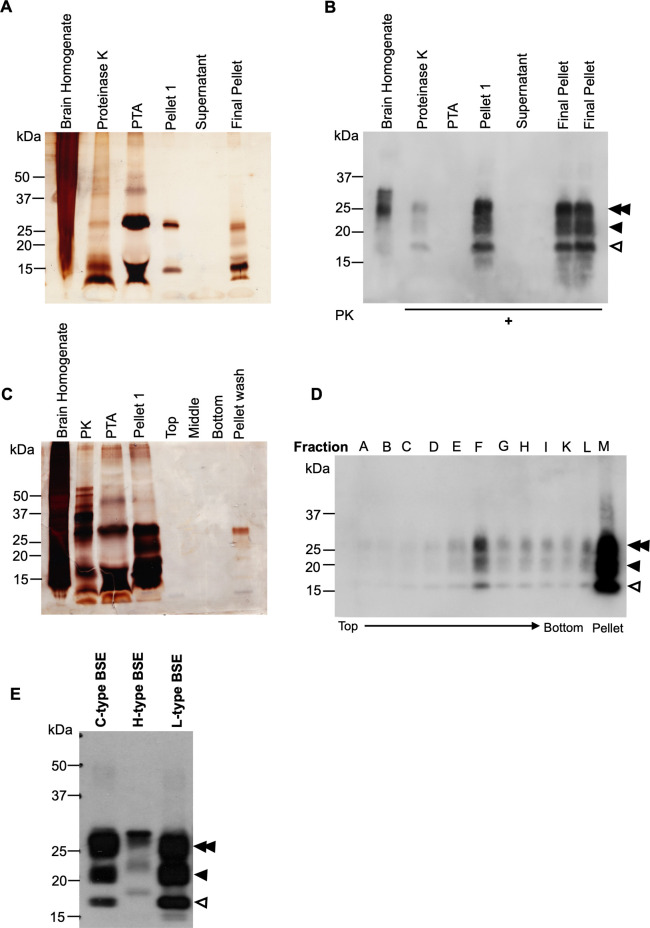
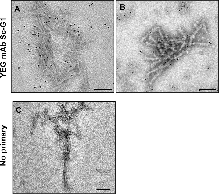
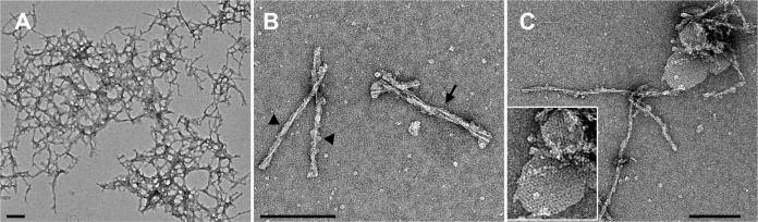
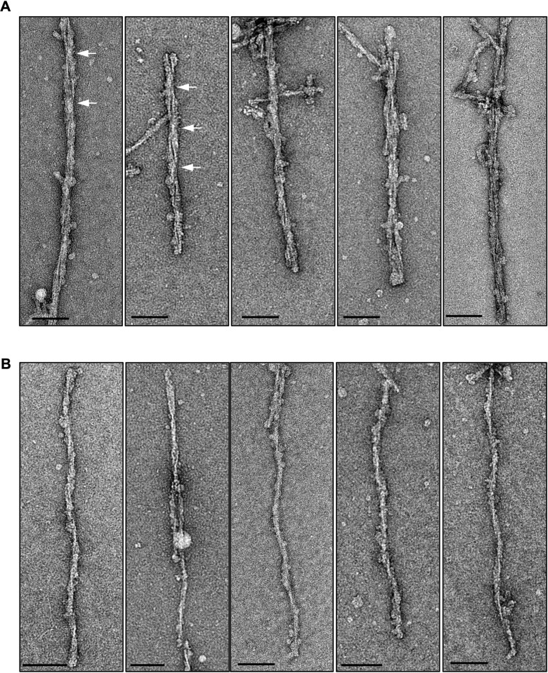
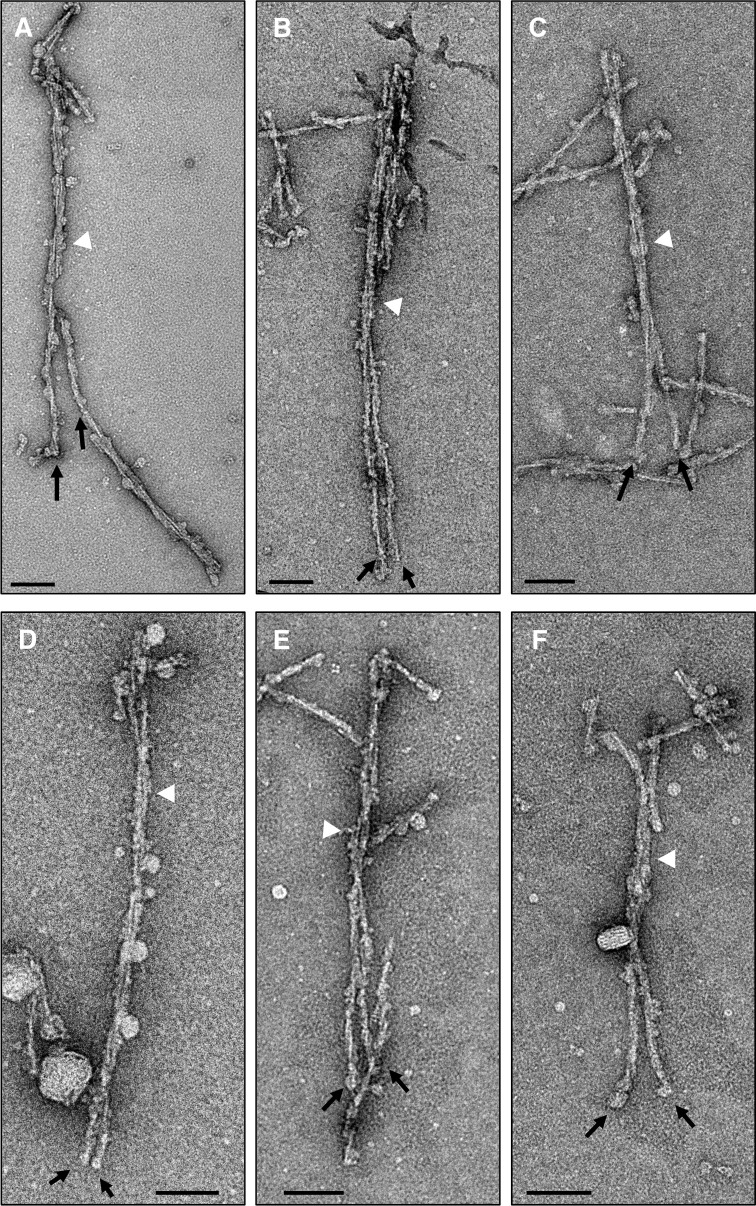
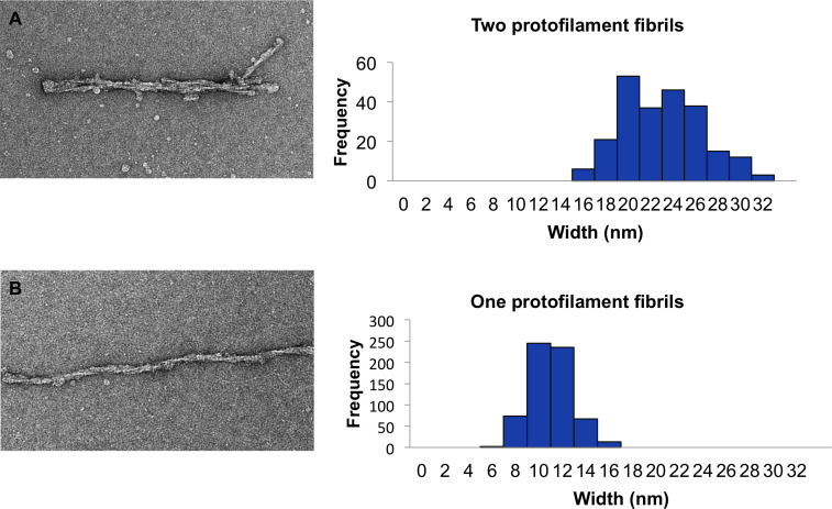
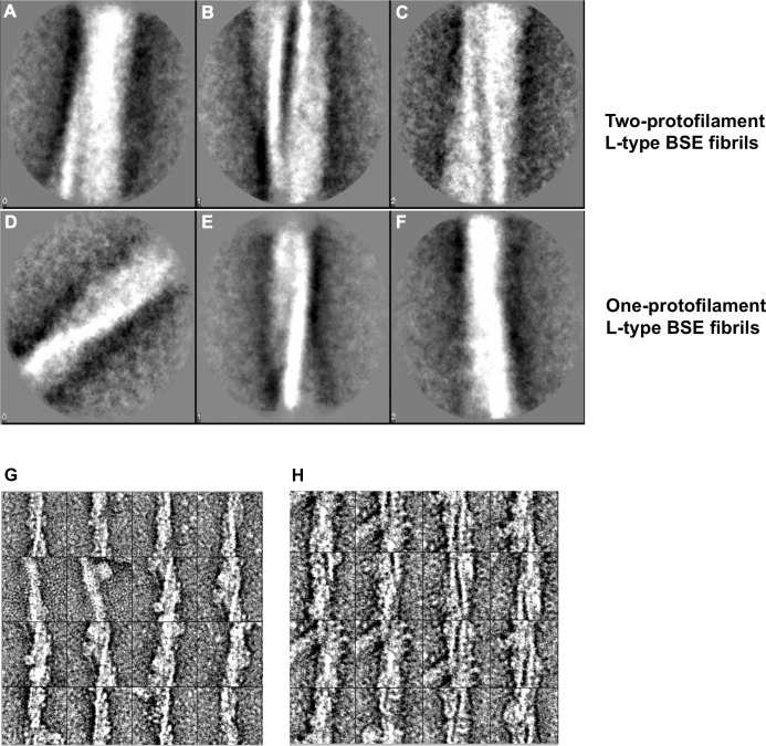
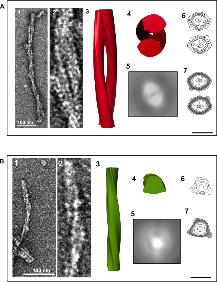
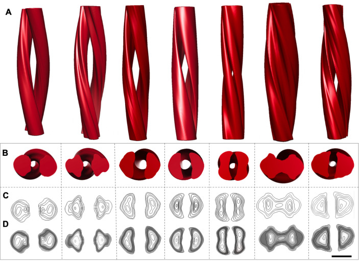
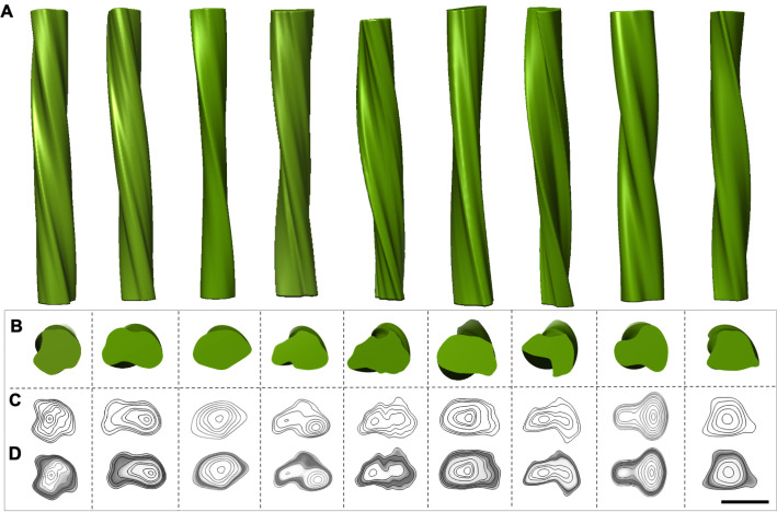
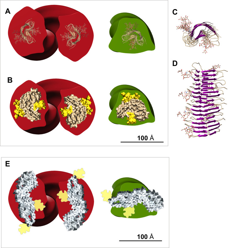
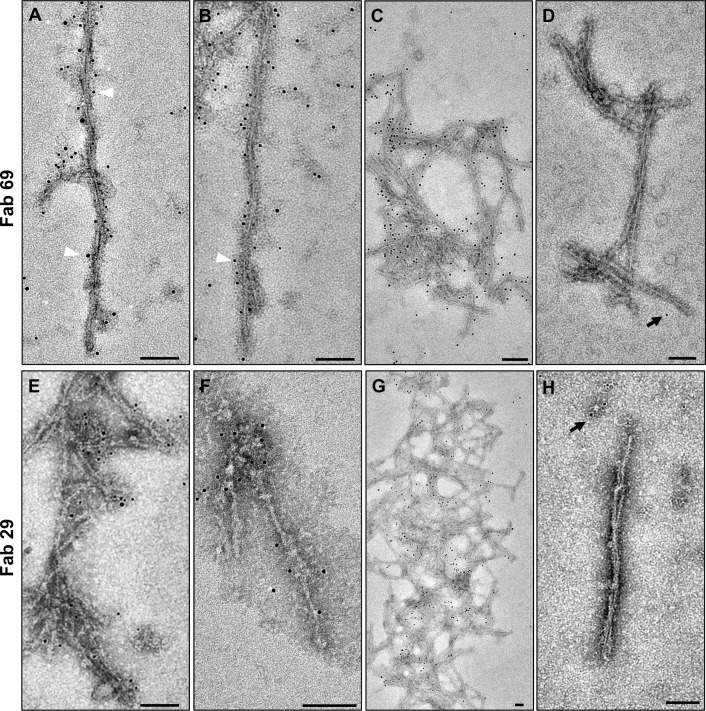
Bovine spongiform encephalopathy (BSE) is a prion disease of cattle that is caused by the misfolding of the cellular prion protein (PrPC) into an infectious conformation (PrPSc). PrPC is a predominantly α-helical membrane protein that misfolds into a β-sheet rich, infectious state, which has a high propensity to self-assemble into amyloid fibrils. Three strains of BSE prions can cause prion disease in cattle, including classical BSE (C-type) and two atypical strains, named L-type and H-type BSE. To date, there is no detailed information available about the structure of any of the infectious BSE prion strains. In this study, we purified L-type BSE prions from transgenic mouse brains and investigated their biochemical and ultrastructural characteristics using electron microscopy, image processing, and immunogold labeling techniques. By using phosphotungstate anions (PTA) to precipitate PrPSc combined with sucrose gradient centrifugation, a high yield of proteinase K-resistant BSE amyloid fibrils was obtained. A morphological examination using electron microscopy, two-dimensional class averages, and three-dimensional reconstructions revealed two structural classes of L-type BSE amyloid fibrils; fibrils that consisted of two protofilaments with a central gap and an average width of 22.5 nm and one-protofilament fibrils that were 10.6 nm wide. The one-protofilament fibrils were found to be more abundant compared to the thicker two-protofilament fibrils. Both fibrillar assemblies were successfully decorated with monoclonal antibodies against N- and C-terminal epitopes of PrP using immunogold-labeling techniques, confirming the presence of polypeptides that span residues 100-110 to 227-237. The fact that the one-protofilament fibrils contain both N- and C-terminal PrP epitopes constrains molecular models for the structure of the infectious conformer in favour of a compact four-rung β-solenoid fold.

摘要

牛海绵状脑病(BSE)是一种牛朊病毒病,由细胞朊病毒蛋白(PrPC)错误折叠成传染性构象(PrPSc)引起。PrPC 是一种主要由α-螺旋膜蛋白组成,错误折叠成富含β-片层的传染性状态,具有高度自我组装成淀粉样纤维的倾向。三种 BSE 朊病毒株可引起牛朊病毒病,包括经典 BSE(C 型)和两种非典型株,命名为 L 型和 H 型 BSE。迄今为止,尚无关于任何传染性 BSE 朊病毒株结构的详细信息。在这项研究中,我们从转基因小鼠脑中纯化了 L 型 BSE 朊病毒,并使用电子显微镜、图像处理和免疫金标记技术研究了它们的生化和超微结构特征。通过使用磷钨酸阴离子(PTA)沉淀 PrPSc 并结合蔗糖梯度离心,获得了高产量的蛋白酶 K 抗性 BSE 淀粉样纤维。使用电子显微镜、二维类平均和三维重建进行形态学检查,揭示了 L 型 BSE 淀粉样纤维的两种结构类型;由两条带有中央间隙的原纤维组成的纤维,平均宽度为 22.5nm,以及宽度为 10.6nm 的单原纤维纤维。与较厚的双原纤维纤维相比,发现单原纤维纤维更为丰富。两种纤维组装体都成功地用针对 PrP N 和 C 末端表位的单克隆抗体进行了免疫金标记,证实了存在跨越残基 100-110 至 227-237 的多肽。单原纤维纤维包含 N 和 C 末端 PrP 表位的事实,有利于支持紧凑的四梯β-发夹折叠的结构模型。

https://cdn.ncbi.nlm.nih.gov/pmc/blobs/ab3b/8195424/73e9dd90e1fe/ppat.1009628.g012.jpg
https://cdn.ncbi.nlm.nih.gov/pmc/blobs/ab3b/8195424/36efd628eac1/ppat.1009628.g001.jpg
https://cdn.ncbi.nlm.nih.gov/pmc/blobs/ab3b/8195424/6f3f410926e8/ppat.1009628.g002.jpg
https://cdn.ncbi.nlm.nih.gov/pmc/blobs/ab3b/8195424/b716fad3cc96/ppat.1009628.g003.jpg
https://cdn.ncbi.nlm.nih.gov/pmc/blobs/ab3b/8195424/dfe66e18ae8b/ppat.1009628.g004.jpg
https://cdn.ncbi.nlm.nih.gov/pmc/blobs/ab3b/8195424/697970b754db/ppat.1009628.g005.jpg
https://cdn.ncbi.nlm.nih.gov/pmc/blobs/ab3b/8195424/8f6447112b5b/ppat.1009628.g006.jpg
https://cdn.ncbi.nlm.nih.gov/pmc/blobs/ab3b/8195424/7d044a8889e7/ppat.1009628.g007.jpg
https://cdn.ncbi.nlm.nih.gov/pmc/blobs/ab3b/8195424/92ac7321ac0a/ppat.1009628.g008.jpg
https://cdn.ncbi.nlm.nih.gov/pmc/blobs/ab3b/8195424/c0b647cdc674/ppat.1009628.g009.jpg
https://cdn.ncbi.nlm.nih.gov/pmc/blobs/ab3b/8195424/336ffd21b03b/ppat.1009628.g010.jpg
https://cdn.ncbi.nlm.nih.gov/pmc/blobs/ab3b/8195424/5450380939e9/ppat.1009628.g011.jpg
https://cdn.ncbi.nlm.nih.gov/pmc/blobs/ab3b/8195424/73e9dd90e1fe/ppat.1009628.g012.jpg

相似文献

1
The ultrastructure of infectious L-type bovine spongiform encephalopathy prions constrains molecular models.传染性 L 型牛海绵状脑病朊病毒的超微结构限制了分子模型。
PLoS Pathog. 2021 Jun 1;17(6):e1009628. doi: 10.1371/journal.ppat.1009628. eCollection 2021 Jun.
2
Transmissibility of H-Type Bovine Spongiform Encephalopathy to Hamster PrP Transgenic Mice.H型牛海绵状脑病对仓鼠朊蛋白转基因小鼠的传染性
PLoS One. 2015 Oct 14;10(10):e0138977. doi: 10.1371/journal.pone.0138977. eCollection 2015.
3
Experimental Infection of Cattle With a Novel Prion Derived From Atypical H-Type Bovine Spongiform Encephalopathy.用源自非典型H型牛海绵状脑病的新型朊病毒对牛进行实验性感染。
Vet Pathol. 2017 Nov;54(6):892-900. doi: 10.1177/0300985817717769. Epub 2017 Jul 21.
4
Absence of Evidence for a Causal Link between Bovine Spongiform Encephalopathy Strain Variant L-BSE and Known Forms of Sporadic Creutzfeldt-Jakob Disease in Human PrP Transgenic Mice.在人类朊蛋白转基因小鼠中,缺乏牛海绵状脑病毒株变体L-BSE与散发性克雅氏病已知形式之间存在因果联系的证据。
J Virol. 2016 Nov 14;90(23):10867-10874. doi: 10.1128/JVI.01383-16. Print 2016 Dec 1.
5
PrP-C1 fragment in cattle brains reveals features of the transmissible spongiform encephalopathy associated PrP.牛脑中的PrP-C1片段揭示了与传染性海绵状脑病相关的朊蛋白的特征。
Brain Res. 2017 Mar 15;1659:19-28. doi: 10.1016/j.brainres.2017.01.015. Epub 2017 Jan 22.
6
Accumulation of mono-glycosylated form-rich, plaque-forming PrPSc in the second atypical bovine spongiform encephalopathy case in Japan.日本第二例非典型牛海绵状脑病病例中富含单糖基化形式、形成斑块的PrPSc的积累。
Jpn J Infect Dis. 2007 Sep;60(5):305-8.
7
The Structural Architecture of an Infectious Mammalian Prion Using Electron Cryomicroscopy.利用电子冷冻显微镜技术解析感染性哺乳动物朊病毒的结构架构
PLoS Pathog. 2016 Sep 8;12(9):e1005835. doi: 10.1371/journal.ppat.1005835. eCollection 2016 Sep.
8
Prion Protein Devoid of the Octapeptide Repeat Region Delays Bovine Spongiform Encephalopathy Pathogenesis in Mice.缺乏八肽重复区域的朊病毒蛋白可延缓小鼠牛海绵状脑病的发病进程。
J Virol. 2017 Dec 14;92(1). doi: 10.1128/JVI.01368-17. Print 2018 Jan 1.
9
Influence of Interspecies Transmission of Atypical Bovine Spongiform Encephalopathy Prions to Hamsters on Prion Characteristics.非典型牛海绵状脑病朊病毒跨物种传播至仓鼠对朊病毒特性的影响
Front Vet Sci. 2020 Mar 3;7:94. doi: 10.3389/fvets.2020.00094. eCollection 2020.
10
Characterization of Syrian hamster adapted prions derived from L-type and C-type bovine spongiform encephalopathies.从 L 型和 C 型牛海绵状脑病分离的叙利亚仓鼠适应朊病毒的特性。
Prion. 2011 Apr-Jun;5(2):103-8. doi: 10.4161/pri.5.2.15847. Epub 2011 Apr 1.

引用本文的文献

1
Bovine spongiform encephalopathy: A review of current knowledge and challenges.牛海绵状脑病:当前知识与挑战综述
Open Vet J. 2025 Jan;15(1):54-68. doi: 10.5455/OVJ.2025.v15.i1.5. Epub 2025 Jan 31.
2
Prion protein E219K polymorphism: from the discovery of the KANNO blood group to interventions for human prion disease.朊病毒蛋白E219K多态性:从神野血型的发现到人类朊病毒病的干预措施
Front Neurol. 2024 Jul 10;15:1392984. doi: 10.3389/fneur.2024.1392984. eCollection 2024.
3
BSE risk posed by ruminant collagen and gelatine derived from bones.

本文引用的文献

1
High-resolution structure and strain comparison of infectious mammalian prions.传染性哺乳动物朊病毒的高分辨率结构和应变比较。
Mol Cell. 2021 Nov 4;81(21):4540-4551.e6. doi: 10.1016/j.molcel.2021.08.011. Epub 2021 Aug 25.
2
Modeling PrP Generation Through Deformed Templating.通过变形模板模拟朊蛋白(PrP)的生成。
Front Bioeng Biotechnol. 2020 Oct 6;8:590501. doi: 10.3389/fbioe.2020.590501. eCollection 2020.
3
Protective anti-prion antibodies in human immunoglobulin repertoires.人类免疫球蛋白库中的保护性抗朊病毒抗体。
源自骨头的反刍动物胶原蛋白和明胶所带来的牛海绵状脑病风险。
EFSA J. 2024 Jul 16;22(7):e8883. doi: 10.2903/j.efsa.2024.8883. eCollection 2024 Jul.
4
Misfolded protein oligomers: mechanisms of formation, cytotoxic effects, and pharmacological approaches against protein misfolding diseases.错误折叠的蛋白质寡聚物:形成机制、细胞毒性作用以及针对蛋白质错误折叠疾病的药理学方法。
Mol Neurodegener. 2024 Feb 20;19(1):20. doi: 10.1186/s13024-023-00651-2.
5
Excess PrP inhibits muscle cell differentiation via miRNA-enhanced liquid-liquid phase separation implicated in myopathy.过量的朊病毒蛋白通过 miRNA 增强的液-液相分离抑制肌肉细胞分化,该过程与肌肉疾病有关。
Nat Commun. 2023 Dec 8;14(1):8131. doi: 10.1038/s41467-023-43826-7.
6
Realization of Amyloid-like Aggregation as a Common Cause for Pathogenesis in Diseases.认识到淀粉样蛋白样聚集是疾病发病机制的常见原因。
Life (Basel). 2023 Jul 7;13(7):1523. doi: 10.3390/life13071523.
7
Inducing prion protein shedding as a neuroprotective and regenerative approach in pathological conditions of the brain: from theory to facts.诱导朊病毒蛋白脱落作为大脑病理状况下的一种神经保护和再生方法:从理论到事实。
Neural Regen Res. 2023 Sep;18(9):1869-1875. doi: 10.4103/1673-5374.366496.
8
Conformation-Dependent Influences of Hydrophobic Amino Acids in Two In-Register Parallel β-Sheet Amyloids, an α-Synuclein Amyloid and a Local Structural Model of PrP.两种平行β-折叠淀粉样蛋白(α-突触核蛋白淀粉样蛋白和朊蛋白局部结构模型)中疏水氨基酸的构象依赖性影响
ACS Omega. 2022 Aug 24;7(35):31271-31288. doi: 10.1021/acsomega.2c03523. eCollection 2022 Sep 6.
9
2.7 Å cryo-EM structure of ex vivo RML prion fibrils.2.7 Å 冷冻电镜结构的 RML 朊病毒纤维。
Nat Commun. 2022 Jul 13;13(1):4004. doi: 10.1038/s41467-022-30457-7.
EMBO Mol Med. 2020 Sep 7;12(9):e12739. doi: 10.15252/emmm.202012739. Epub 2020 Aug 10.
4
Cryo-EM structure of an amyloid fibril formed by full-length human prion protein.全长人类朊病毒蛋白形成的淀粉样纤维的冷冻电镜结构。
Nat Struct Mol Biol. 2020 Jun;27(6):598-602. doi: 10.1038/s41594-020-0441-5. Epub 2020 Jun 8.
5
Isolation of infectious, non-fibrillar and oligomeric prions from a genetic prion disease.从遗传性朊病毒病中分离传染性、无纤维和寡聚朊病毒。
Brain. 2020 May 1;143(5):1512-1524. doi: 10.1093/brain/awaa078.
6
Full atomistic model of prion structure and conversion.朊病毒结构和转化的全原子模型。
PLoS Pathog. 2019 Jul 11;15(7):e1007864. doi: 10.1371/journal.ppat.1007864. eCollection 2019 Jul.
7
Structural features distinguishing infectious ex vivo mammalian prions from non-infectious fibrillar assemblies generated in vitro.将感染性哺乳动物朊病毒与体外生成的无感染性纤维状组装体区分开来的结构特征。
Sci Rep. 2019 Jan 23;9(1):376. doi: 10.1038/s41598-018-36700-w.
8
The prion 2018 round tables (I): the structure of PrP.朊病毒2018年圆桌会议(一):朊蛋白的结构
Prion. 2019 Jan;13(1):46-52. doi: 10.1080/19336896.2019.1569450.
9
Comparing the Folds of Prions and Other Pathogenic Amyloids.朊病毒与其他致病性淀粉样蛋白的折叠比较
Pathogens. 2018 May 4;7(2):50. doi: 10.3390/pathogens7020050.
10
Prion infectivity is encoded exclusively within the structure of proteinase K-resistant fragments of synthetically generated recombinant PrP.朊病毒感染性完全由合成产生的重组朊蛋白蛋白酶 K 抗性片段的结构编码。
Acta Neuropathol Commun. 2018 Apr 24;6(1):30. doi: 10.1186/s40478-018-0534-0.