Translational Imaging Center, Houston Methodist Research Institute, Houston, Texas, USA.
Department of Urology, Houston Methodist Hospital, Houston, Texas, USA.
Neurourol Urodyn. 2022 Sep;41(7):1612-1619. doi: 10.1002/nau.25008. Epub 2022 Jul 17.
A number of neurourology imaging studies have mainly focused on investigating the brain activations during micturition in healthy and neuropathic patients. It is, however, also necessary to study brain functional connectivity (FC) within bladder-related regions to understand the brain organization during the execution of bladder function. This study aims to identify the altered brain network associated with bladder function in multiple sclerosis (MS) women with voiding dysfunction through comparisons with healthy subjects via concurrent urodynamic study (UDS)/functional magnetic resonance imaging (fMRI).
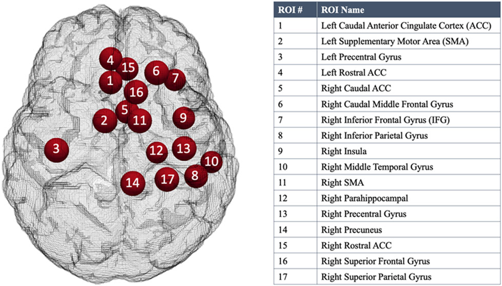
Ten healthy adult women and nine adult ambulatory women with clinically stable MS for ≥6 months and symptomatic voiding phase neurogenic lower urinary tract dysfunction (NLUTD) underwent UDS/fMRI evaluation with a task of bladder filling/emptying that was repeated three to five times. We quantitatively compared their FC within 17 bladder-related brain regions during two UDS phases: "strong desire to void" and "(attempt at) voiding initiation."
At "strong desire to void," the healthy group showed significantly stronger FC in regions involved in bladder filling and suppression of voiding compared to the patient group. These regions included the bilateral anterior cingulate cortex, right supplementary motor area, and right middle frontal gyrus. During "(attempt at) voiding initiation," healthy subjects exhibited stronger FC in the right inferior frontal gyrus compared to MS patients.
Our study offers a new way to identify alterations in the neural mechanisms underlying NLUTD and provides potential targets for clinical interventions (such as cortical neuromodulation) aimed at restoring bladder functions in MS patients.
许多神经泌尿影像学研究主要集中在研究健康和神经病变患者排尿过程中的大脑激活。然而,研究与膀胱相关区域的大脑功能连接(FC)对于理解膀胱功能执行过程中的大脑组织也是必要的。本研究旨在通过与健康受试者的同步尿动力学研究/功能磁共振成像(fMRI)比较,确定多发性硬化症(MS)女性排尿功能障碍患者与膀胱功能相关的改变脑网络。
10 名健康成年女性和 9 名有临床稳定 MS 病史且有症状性排尿期神经原性下尿路功能障碍(NLUTD)的成年活动女性接受了 UDS/fMRI 评估,评估时进行了膀胱充盈/排空任务,重复 3 到 5 次。我们在两个 UDS 阶段(“强烈排尿欲望”和“(尝试)排尿起始”)比较了 17 个与膀胱相关的大脑区域内的他们的 FC。
在“强烈排尿欲望”时,与患者组相比,健康组在涉及膀胱充盈和抑制排尿的区域中显示出明显更强的 FC。这些区域包括双侧前扣带皮质、右侧辅助运动区和右侧额中回。在“(尝试)排尿起始”期间,与 MS 患者相比,健康受试者在右侧额下回显示出更强的 FC。
我们的研究提供了一种识别 NLUTD 下神经机制改变的新方法,并为旨在恢复 MS 患者膀胱功能的临床干预(如皮质神经调节)提供了潜在靶点。