Clinic of Gastroenterology and Hepatology, Clinical Center of Serbia, Belgrade, Serbia.
Faculty of Medicine, University of Belgrade, Belgrade, Serbia.
Int J Clin Pract. 2022 Jul 9;2022:3339866. doi: 10.1155/2022/3339866. eCollection 2022.
Ultrasonography is a noninvasive, inexpensive, and widely available diagnostic tool. In the last two decades, the development of ultrasound techniques and equipment has significantly increased the usage of intestine ultrasound (US) in the assessment of the gastrointestinal tract in patients with inflammatory bowel disease (IBD). Although current guidelines suggest routine utilization of US in patients with Crohn's disease, data regarding US usage in ulcerative colitis are still scarce. We aimed to assess the reliability of intestinal ultrasonography in the assessment of disease activity and extension of patients with ulcerative colitis.
Fifty-five patients with a histologically confirmed diagnosis of ulcerative colitis, treated at University Clinical Center of Serbia in the period from 2019 to 2022 were included in this retrospective observational study. The data were obtained from the patient's medical records including history, laboratory, US, and endoscopy findings. US examined parameters were as following: bowel wall thickness (BWT), presence of fat wrapping, wall layer stratification, mesenteric hypertrophy, presence of enlarged mesenteric lymph nodes, and absence or presence of ascites.
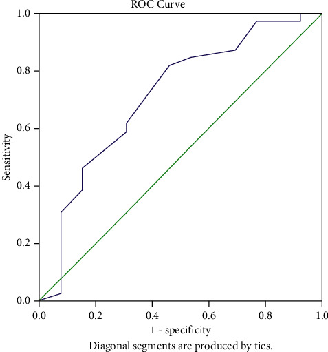
Our results suggest that there is a strong correlation of BWT and colonoscopy findings regarding disease extension ( = 0.524, =0.01, < 0.05). Furthermore, our results have shown a statistically significant correlation of BWT with the Mayo endoscopic score ( = 0.434, =0.01, < 0.05), disease activity score ( = 0.369,=0.01, < 0.05), degree of ulcerative colitis burden of luminal inflammation ( = 0.366, =0.01, < 0.05), and Geboes index ( = 0.298, =0.027, < 0.05). Overall accuracy of US for disease extension and activity was statistically significant ( < 0.05).
Our results suggest that US is a moderately accurate method for the assessment of disease activity and localization in patients with UC.
超声检查是一种非侵入性、廉价且广泛应用的诊断工具。在过去的二十年中,超声技术和设备的发展显著增加了肠超声(US)在炎症性肠病(IBD)患者胃肠道评估中的应用。尽管目前的指南建议在克罗恩病患者中常规使用 US,但关于溃疡性结肠炎中 US 使用的数据仍然很少。我们旨在评估肠超声在溃疡性结肠炎患者疾病活动度和范围评估中的可靠性。
本回顾性观察研究纳入了 2019 年至 2022 年期间在塞尔维亚大学临床中心接受组织学确诊溃疡性结肠炎治疗的 55 例患者。数据来自患者病历,包括病史、实验室、US 和内镜检查结果。US 检查的参数如下:肠壁厚度(BWT)、脂肪包裹的存在、壁层分层、肠系膜肥厚、肠系膜淋巴结肿大、腹水的存在或不存在。
我们的结果表明,BWT 与结肠镜检查结果在疾病范围上具有较强的相关性( = 0.524,=0.01,<0.05)。此外,我们的结果还显示 BWT 与 Mayo 内镜评分( = 0.434,=0.01,<0.05)、疾病活动评分( = 0.369,=0.01,<0.05)、溃疡性结肠炎黏膜炎症负担程度( = 0.366,=0.01,<0.05)和 Geboes 指数( = 0.298,=0.027,<0.05)之间存在统计学显著相关性。US 对疾病范围和活动的整体准确性具有统计学意义(<0.05)。
我们的结果表明,US 是一种评估 UC 患者疾病活动度和定位的中等准确性方法。