Nakazawa Takahiko, Aihara Masanori, Mizuno Hiroyuki, Yamaguchi Rei, Yoshimoto Yuhei
Department of Neurosurgery, Gunma University Graduate School of Medicine, Mebashi.
Department of Neurosurgery, Subaru Health Insurance Society Ota Memorial Hospital, Ota, Gunma, Japan.
Surg Neurol Int. 2022 Jun 23;13:275. doi: 10.25259/SNI_95_2022. eCollection 2022.
Meningioma and dural arteriovenous fistula (dAVF) located at the same site are rare. The present case demonstrated the transformation of tumor feeding vessels into the pial feeder of the dAVF over time, which may help to elucidate the pathogenesis of tumor-associated dAVF.
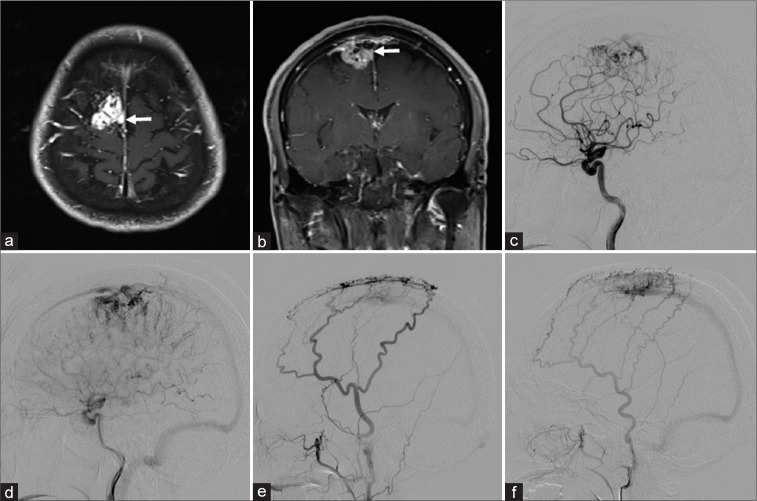
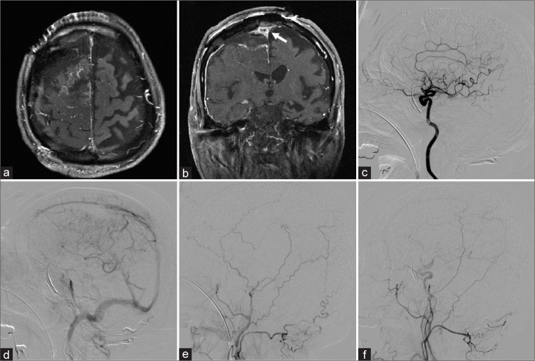
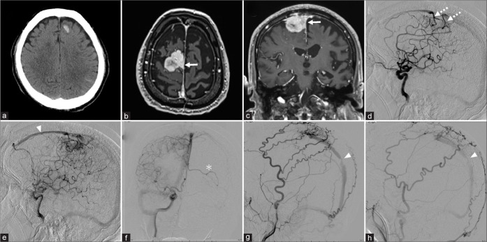
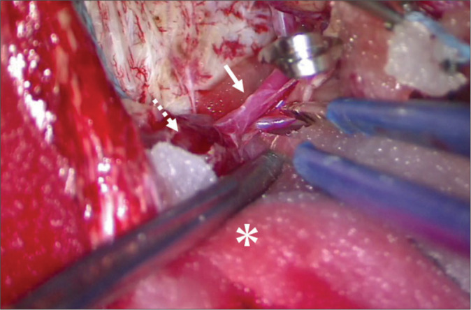
A 71-year-old man presented with convulsion. Magnetic resonance (MR) imaging showed a right parasagittal sinus meningioma invading the superior sagittal sinus (SSS). Bilateral external carotid angiography showed dAVF at the SSS, near the site of tumor invasion. The right internal carotid angiography showed tumor staining from the anterior cerebral artery with intra-tumor arteriovenous shunting, with stagnation of tumor blood flow, suggesting impairment of perfusion to the SSS. Four years after the initial diagnosis, the patient was admitted to hospital with status epilepticus, and MR imaging showed an enlarged tumor. Carotid angiography revealed transformation of the tumor feeders to the pial feeder of the dAVF. The findings of shunting to the SSS had intensified, and stenosis had occurred in the posterior third of the SSS. The venous return showed retrograde flow anteriorly to the SSS. The patient underwent endovascular embolization and tumor resection. The shunt had disappeared.
This report supports the proposal that impaired venous return is an important factor in the shunt occurrence of dAVF. Neurosurgeons should consider that cases of meningioma invading the venous sinuses may be complicated by dAVF and changes may occur over time.
位于同一部位的脑膜瘤和硬脑膜动静脉瘘(dAVF)较为罕见。本病例展示了肿瘤供血血管随时间转变为dAVF的软膜供血血管,这可能有助于阐明肿瘤相关性dAVF的发病机制。
一名71岁男性因惊厥就诊。磁共振(MR)成像显示右侧矢状窦旁脑膜瘤侵犯上矢状窦(SSS)。双侧颈外动脉血管造影显示在肿瘤侵犯部位附近的SSS处存在dAVF。右侧颈内动脉血管造影显示大脑前动脉有肿瘤染色且肿瘤内存在动静脉分流,肿瘤血流停滞,提示SSS灌注受损。初次诊断4年后,患者因癫痫持续状态入院,MR成像显示肿瘤增大。颈动脉血管造影显示肿瘤供血血管转变为dAVF的软膜供血血管。向SSS分流的情况加剧,且SSS后三分之一处出现狭窄。静脉回流显示向前逆行至SSS。患者接受了血管内栓塞和肿瘤切除术。分流消失。
本报告支持静脉回流受损是dAVF分流发生的重要因素这一观点。神经外科医生应考虑到脑膜瘤侵犯静脉窦的病例可能并发dAVF,且情况可能随时间发生变化。