Ngo Khoa Xuan, Vo Huy Tien, Nguyen Duong Thai-Ha, Doan Ha Thi-Ngoc
Hanoi Medical University, Viet Nam.
Hong Ha General Hospital, Viet Nam.
Ann Med Surg (Lond). 2022 Jun 17;79:103996. doi: 10.1016/j.amsu.2022.103996. eCollection 2022 Jul.
Both free medial sural artery perforator flaps and pedicled medial sural artery perforator flaps have been being effectively used in treatment of body defects especially in head and neck region by plastic surgeons worldwide. However, there is a lack of comprehensive studies on the anatomy of perforating artery branches in Vietnam. This study aims to describe anatomical vascular pedicles of medial sural artery perforator flap in Vietnamese adults
A descriptive cross-sectional study, dissected 62 lower limbs of 41 Vietnamese adult cadavers preserved by formalin in Department of Anatomy, Hanoi Medical University and Ho Chi Minh Medicine and Pharmacy University.
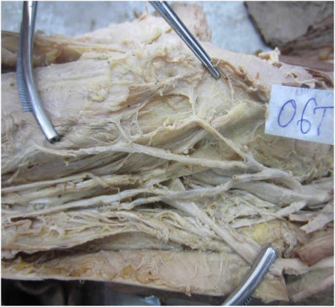
Origin of medial sural artery was branched constantly from popliteal artery. Common stem of artery was 8.39 ± 3.5 cm in mean length. The diameter of common stem, which was measured from origin, was 2.88 ± 0.98 mm averagely. The common stem of artery, which did not have any branch (15%), divided in to 2 branches (15%), 3 branches (30%), 4 branches (40%) before entering muscle. Medial sural artery had 1 to 5 branches perforating to the skin. The distance from perforating branch to the knee joint (popliteal crease) was 10.12 ± 3.7 cm, the distance from perforator branch to middle posterior leg was 1.6 ± 0.96 cm
The medial sural artery constantly originates from popliteal artery, supplies blood for medial gastrocnemius muscle. The skin area covering this muscle is nourished by one of five perforators of the medial sural artery. The perforating flaps can be created using medial sural artery perforating branches.
游离腓肠内侧动脉穿支皮瓣和带蒂腓肠内侧动脉穿支皮瓣均已被全球整形外科医生有效地用于治疗身体缺损,尤其是头颈部缺损。然而,越南缺乏关于穿支动脉分支解剖的全面研究。本研究旨在描述越南成年人腓肠内侧动脉穿支皮瓣的解剖血管蒂。
一项描述性横断面研究,解剖了河内医科大学和胡志明医药大学解剖学系用福尔马林保存的41具越南成年尸体的62条下肢。
腓肠内侧动脉起源恒定于腘动脉。动脉总干平均长度为8.39±3.5厘米。从起源处测量的总干直径平均为2.88±0.98毫米。动脉总干无分支的占15%,进入肌肉前分为2支的占15%,3支的占30%,4支的占40%。腓肠内侧动脉有1至5支穿支至皮肤。穿支至膝关节(腘横纹)的距离为10.12±3.7厘米,穿支至小腿中后部的距离为1.6±0.96厘米。
腓肠内侧动脉恒定起源于腘动脉,为腓肠肌内侧头供血。覆盖该肌肉的皮肤区域由腓肠内侧动脉的五支穿支之一供血。可使用腓肠内侧动脉穿支创建穿支皮瓣。