Department of Diagnostic and Interventional Radiology, University Hospital RWTH Aachen, Pauwelsstraße 30, 52074, Aachen, Germany.
Philips Healthcare, Hamburg, Germany.
Sci Rep. 2022 Jul 21;12(1):12468. doi: 10.1038/s41598-022-16324-x.
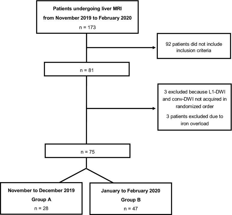
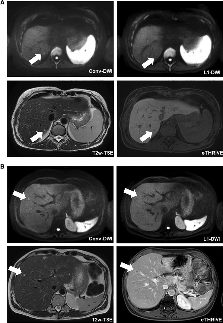
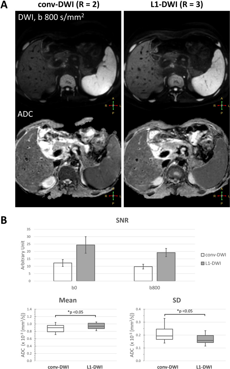
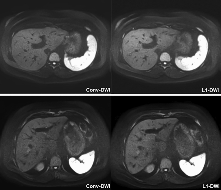
To investigate whether combining L1-regularized iterative sensitivity encoding (SENSE) reconstruction and single-shot echo planar imaging (EPI) is useful in hepatic DWI. Single-shot EPI-DWI with L1-regularized iterative SENSE reconstruction (L1-DWI) and conventional parallel imaging-based reconstruction (conv-DWI) in liver MRI were compared in volunteers and patients. For the patient cohort, 75 subjects (60 ± 13 years) with 349 focal liver lesions (FLL) were included. Patient groups A and B were used to reduce acquisition time or improve spatial resolution, respectively. Image parameters were rated on a 5-point scale. The number of FLLs was recorded; in case of discrepancy, the reason for non-detectability was analyzed. In volunteers, higher signal-to-noise ratio (24.4 ± 5.6 vs. 12.2 ± 2.3, p < 0.001 at b = 0; 19.3 ± 2.8 vs. 9.8 ± 1.6, p < 0.001 at b = 800) and lower standard deviation of the apparent diffusion coefficient-values (0.17 vs. 0.20 mm/s, p < 0.05) were found on L1-DWI compared to conv-DWI. In patients, image ratings were similar for all parameters except for "conspicuity of FLLs" which was rated significantly lower on L1-DWI vs. conv-DWI (4.7 ± 0.6 vs. 4.2 ± 0.9, p < 0.05) in group A. In five patients, 11/349 FLLs were not detectable on L1-DWI, but on conv-DWI. L1-regularized iterative reconstruction of single-shot EPI DWI can accelerate image acquisition or improve spatial resolution. However, our finding that FLLs were non-detectable on L1-DWI warrants further research.
为了探究 L1 正则化迭代敏感编码(SENSE)重建与单次回波平面成像(EPI)联合应用于肝脏弥散加权成像(DWI)是否具有优势,我们对比了志愿者和患者肝脏 MRI 中单次激发 EPI-DWI 联合 L1 正则化迭代 SENSE 重建(L1-DWI)和传统并行成像重建(conv-DWI)的结果。在患者队列中,我们纳入了 75 例(60±13 岁)患者,共计 349 个局灶性肝脏病变(FLL)。患者分组 A 和 B 分别用于缩短采集时间和提高空间分辨率。通过 5 分制评分对图像参数进行了评价。记录了 FLL 的数量,对于未检测到的 FLL,我们分析了原因。在志愿者中,L1-DWI 组的信噪比(24.4±5.6 比 12.2±2.3,b=0 时 p<0.001;19.3±2.8 比 9.8±1.6,b=800 时 p<0.001)更高,表观弥散系数值的标准差更低(0.17 比 0.20 mm/s,p<0.05)。在患者中,除了“FLL 显示程度”(分组 A 中 L1-DWI 评分 4.7±0.6,conv-DWI 评分 4.2±0.9,p<0.05)外,所有参数的图像评分均相似。在 5 例患者中,11/349 个 FLL 在 L1-DWI 上无法检测到,但在 conv-DWI 上可以。单次激发 EPI DWI 的 L1 正则化迭代重建可以加速图像采集或提高空间分辨率。然而,我们发现 FLL 在 L1-DWI 上无法检测到,这需要进一步研究。