The First Clinical Medical College of Lanzhou University, Lanzhou, China.
Department of Cardiology, The First Hospital of Lanzhou University, No. 1, Donggang West Road, Chengguan District, Lanzhou, 730000, Gansu, China.
BMC Cardiovasc Disord. 2022 Jul 23;22(1):327. doi: 10.1186/s12872-022-02772-w.
Traditional angiography only displays two-dimensional images of the coronary arteries during stent implantation. However, intravascular imaging can show the structure of the vascular wall, plaque characteristics. This article aims to evaluate the efficacy of intravascular imaging-guided drug-eluting stent (DES) implantation.
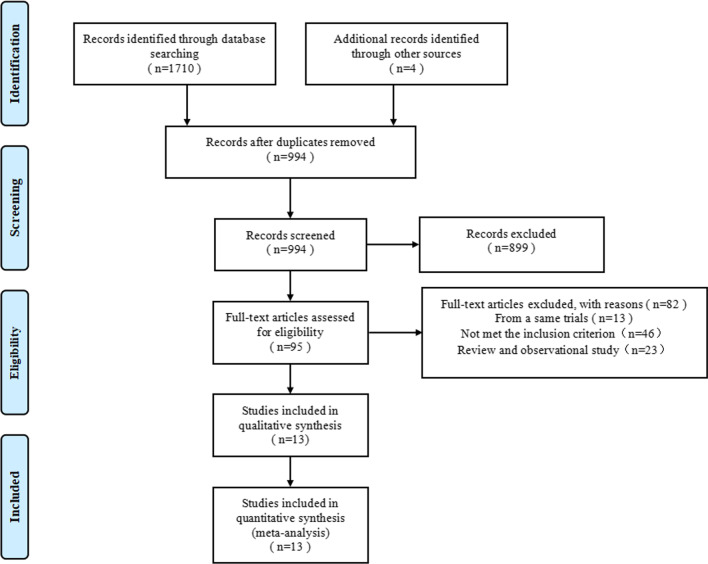
We conducted a systematic review and meta-analysis of randomized controlled trials of intravascular imaging-guided, including patients with DES implantation guided by intravascular ultrasound or optical coherence tomography and traditional angiography. The databases of PubMed, EMBASE, web of science, and Cochrane Library were searched. The primary outcome was target lesion revascularization (TLR). The secondary outcomes included the target vessel revascularization (TVR), myocardial infarction (MI), stent thrombosis (ST), cardiac death, all-cause death, and the major adverse cardiac events (MACE) during the 6-24 months follow-up. The fixed-effects model was used to calculate the relative risk (RR) and 95% confidence interval of the outcome event. Meanwhile, the trial sequence analysis was employed to evaluate the results.
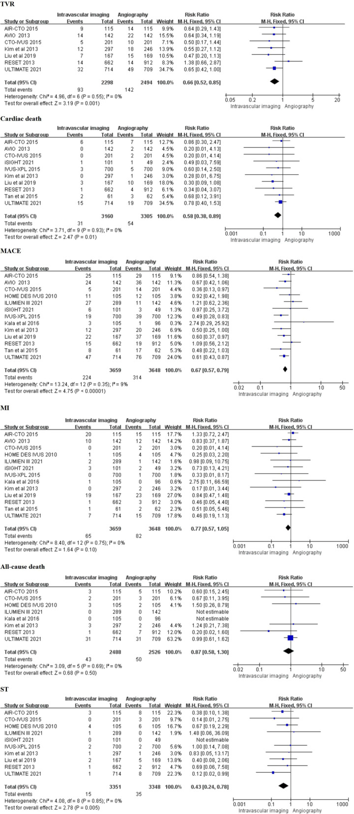
This meta-analysis included fourteen randomized controlled trials with 7307 patients. Compared with angiography-guided, intravascular imaging-guided DES implantation can significantly reduce the risk of TLR (RR 0.63, 0.49-0.82, P = 0.0004), TVR (RR 0.66, 0.52-0.85, P = 0.001), cardiac death (RR 0.58; 0.38-0.89; P = 0.01), MACE (RR 0.67, 0.57-0.79; P < 0.00001) and ST (RR 0.43, 0.24-0.78; P = 0.005). While there was no significant difference regarding MI (RR 0.77, 0.57-1.05, P = 0.10) and all-cause death (RR 0.87, 0.58-1.30, P = 0.50).
Compared with angiography, intravascular imaging-guided DES implantation is associated with better clinical outcomes in patients with coronary artery disease, especially complex lesions (Registered by PROSPERO, CRD 42021289205).
传统血管造影术仅在支架植入过程中显示冠状动脉的二维图像。然而,血管内成像可以显示血管壁的结构、斑块特征。本文旨在评估血管内成像指导下药物洗脱支架(DES)植入的疗效。
我们对血管内成像指导的随机对照试验进行了系统评价和荟萃分析,包括血管内超声或光相干断层扫描指导的 DES 植入患者和传统血管造影指导的患者。检索了 PubMed、EMBASE、Web of Science 和 Cochrane Library 数据库。主要结局为靶病变血运重建(TLR)。次要结局包括靶血管血运重建(TVR)、心肌梗死(MI)、支架血栓形成(ST)、心脏死亡、全因死亡和 6-24 个月随访期间的主要不良心脏事件(MACE)。采用固定效应模型计算结局事件的相对风险(RR)和 95%置信区间。同时,采用试验序列分析评估结果。
这项荟萃分析纳入了 14 项随机对照试验,共 7307 例患者。与血管造影指导相比,血管内成像指导的 DES 植入可显著降低 TLR(RR 0.63,0.49-0.82,P=0.0004)、TVR(RR 0.66,0.52-0.85,P=0.001)、心脏死亡(RR 0.58;0.38-0.89;P=0.01)、MACE(RR 0.67,0.57-0.79;P<0.00001)和 ST(RR 0.43,0.24-0.78;P=0.005)的风险。而 MI(RR 0.77,0.57-1.05,P=0.10)和全因死亡(RR 0.87,0.58-1.30,P=0.50)差异无统计学意义。
与血管造影相比,血管内成像指导的 DES 植入与冠心病患者更好的临床结局相关,特别是复杂病变(由 PROSPERO 注册,CRD42021289205)。