Naumenko Victor A, Vishnevskiy Daniil A, Stepanenko Aleksei A, Sosnovtseva Anastasiia O, Chernysheva Anastasiia A, Abakumova Tatiana O, Valikhov Marat P, Lipatova Anastasiia V, Abakumov Maxim A, Chekhonin Vladimir P
V. Serbsky National Medical Research Center for Psychiatry and Narcology, 119034 Moscow, Russia.
Department of Medical Nanobiotechnology, N.I Pirogov Russian National Research Medical University, 117997 Moscow, Russia.
Biomedicines. 2022 Jul 13;10(7):1697. doi: 10.3390/biomedicines10071697.
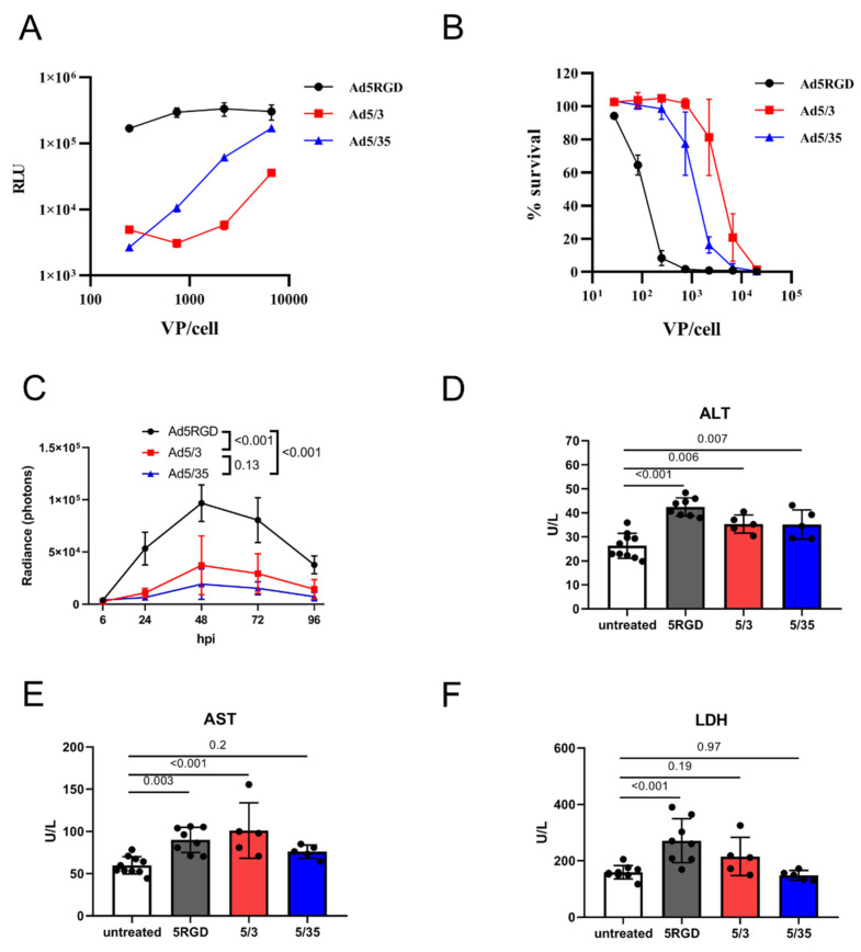
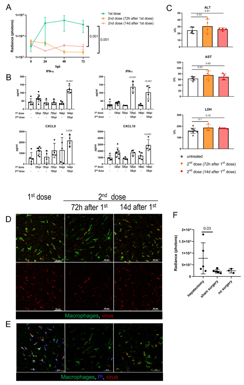
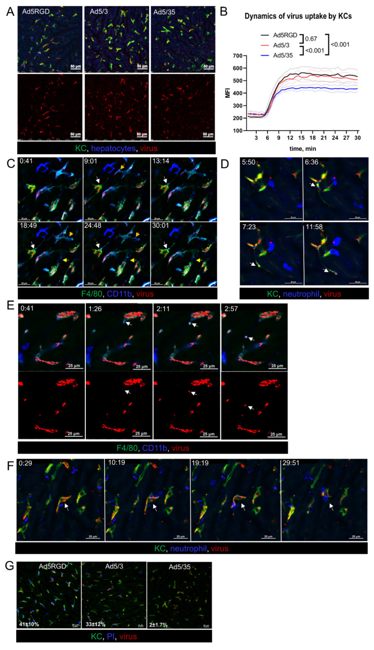
Hepatotoxicity remains an as yet unsolved problem for adenovirus (Ad) cancer therapy. The toxic effects originate both from rapid Kupffer cell (KCs) death (early phase) and hepatocyte transduction (late phase). Several host factors and capsid components are known to contribute to hepatotoxicity, however, the complex interplay between Ad and liver cells is not fully understood. Here, by using intravital microscopy, we aimed to follow the infection and immune response in mouse liver from the first minutes up to 72 h post intravenous injection of three Ads carrying delta-24 modification (Ad5-RGD, Ad5/3, and Ad5/35). At 15-30 min following the infusion of Ad5-RGD and Ad5/3 (but not Ad5/35), the virus-bound macrophages demonstrated signs of zeiosis: the formation of long-extended protrusions and dynamic membrane blebbing with the virus release into the blood in the membrane-associated vesicles. Although real-time imaging revealed interactions between the neutrophils and virus-bound KCs within minutes after treatment, and long-term contacts of CD8+ T cells with transduced hepatocytes at 24-72 h, depletion of neutrophils and CD8+ T cells affected neither rate nor dynamics of liver infection. Ad5-RGD failed to complete replicative cycle in hepatocytes, and transduced cells remained impermeable for propidium iodide, with a small fraction undergoing spontaneous apoptosis. In Ad5-RGD-immune mice, the virus neither killed KCs nor transduced hepatocytes, while in the setting of hepatic regeneration, Ad5-RGD enhanced liver transduction. The clinical and biochemical signs of hepatotoxicity correlated well with KC death, but not hepatocyte transduction. Real-time in vivo tracking for dynamic interactions between virus and host cells provides a better understanding of mechanisms underlying Ad-related hepatotoxicity.
肝毒性仍然是腺病毒(Ad)癌症治疗中一个尚未解决的问题。毒性作用既源于库普弗细胞(KCs)的快速死亡(早期阶段),也源于肝细胞转导(晚期阶段)。已知几种宿主因素和衣壳成分会导致肝毒性,然而,Ad与肝细胞之间复杂的相互作用尚未完全了解。在这里,我们通过活体显微镜检查,旨在追踪静脉注射三种携带delta-24修饰的Ad(Ad5-RGD、Ad5/3和Ad5/35)后,小鼠肝脏从最初几分钟到72小时的感染和免疫反应。在输注Ad5-RGD和Ad5/3(但不是Ad5/35)后的15 - 30分钟,与病毒结合的巨噬细胞出现了细胞溶解的迹象:形成长而延伸的突起以及动态的膜泡形成,病毒通过膜相关囊泡释放到血液中。尽管实时成像显示治疗后几分钟内中性粒细胞与病毒结合的KCs之间存在相互作用,以及24 - 72小时时CD8 + T细胞与转导的肝细胞之间存在长期接触,但中性粒细胞和CD8 + T细胞的耗竭既不影响肝脏感染的速率也不影响其动态过程。Ad5-RGD未能在肝细胞中完成复制周期,转导的细胞对碘化丙啶仍不可渗透,只有一小部分细胞发生自发凋亡。在Ad5-RGD免疫的小鼠中,病毒既不杀死KCs也不转导肝细胞,而在肝再生的情况下,Ad5-RGD增强了肝脏转导。肝毒性的临床和生化体征与KCs死亡密切相关,但与肝细胞转导无关。对病毒与宿主细胞之间动态相互作用的实时体内追踪有助于更好地理解Ad相关肝毒性的潜在机制。