Bao Zheming, Chen Mengli, Li Chen, Shan Qing, Wang Yichen, Yang Wenshan
Department of Pharmacy, Medical Supplies Centre of PLA General Hospital, No. 28, Fuxing Road, Haidian District, Beijing 100853, China.
Orthopedics Department, 960th Hospital of PLA Joint Service Support Force, Jinan, China.
Open Life Sci. 2022 Jul 16;17(1):781-793. doi: 10.1515/biol-2022-0079. eCollection 2022.
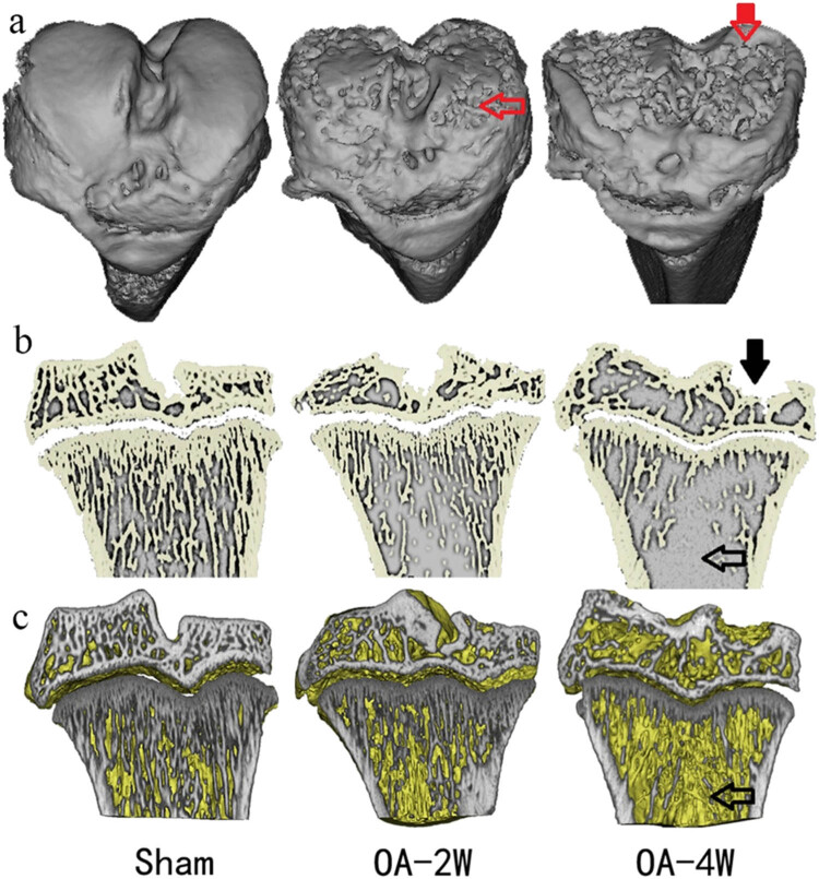
The monosodium iodoacetate (MIA)-induced osteoarthritis (OA) may lead to cartilage degeneration and histopathological lesions. However, the correlation between inflammatory reaction and subchondral bone remodeling in a rodent osteoarthritic model is ambiguous. In this study, intra-articular injection of MIA was performed in 36 four-week-old specific pathogen-free male Wistar rats to induce OA. After 4 weeks of intervention, changes in intrinsic structural properties of the subchondral bones were measured, and the histological evaluation, as well as biochemical analysis, was conducted. We found that intra-articular injection of MIA increased chondrocyte apoptosis and promoted cartilage matrix degradation, such as cartilage surface defects and shallow or disappearing staining. MIA also induced inflammation, improved the expression of IL-1β, TNF-α, and matrix metalloproteinase, and decreased the expression of cartilage-specific proteins with the extension of modeling time. Meanwhile, the MIA also significantly accelerated the subchondral bone remodeling, as shown by the decreased subchondral bone density, thinning of trabeculae, disordered cartilage structure, and morphology. In conclusion, we have shown that MIA-induced rodent osteoarthritic model would cause decreased subchondral bone density, sparse trabecular bone, and other manifestations of osteoporosis accompanied by an inflammatory response, which would worsen with the progression of modeling time. Our results suggest that different phases of MIA-induced OA are associated with the changes in subchondral bone microstructure and the progression of local inflammation.
碘乙酸钠(MIA)诱导的骨关节炎(OA)可能导致软骨退变和组织病理学损伤。然而,在啮齿动物骨关节炎模型中,炎症反应与软骨下骨重塑之间的相关性尚不明确。在本研究中,对36只4周龄的无特定病原体雄性Wistar大鼠进行关节腔内注射MIA以诱导OA。干预4周后,测量软骨下骨的内在结构特性变化,并进行组织学评估和生化分析。我们发现,关节腔内注射MIA增加了软骨细胞凋亡并促进了软骨基质降解,如软骨表面缺损以及染色变浅或消失。随着建模时间的延长,MIA还诱导了炎症反应,提高了白细胞介素-1β(IL-1β)、肿瘤坏死因子-α(TNF-α)和基质金属蛋白酶的表达,并降低了软骨特异性蛋白的表达。同时,MIA还显著加速了软骨下骨重塑,表现为软骨下骨密度降低、小梁变薄、软骨结构和形态紊乱。总之,我们表明MIA诱导的啮齿动物骨关节炎模型会导致软骨下骨密度降低、小梁骨稀疏以及其他骨质疏松表现,并伴有炎症反应,且随着建模时间的推移会恶化。我们的结果表明,MIA诱导的OA的不同阶段与软骨下骨微观结构的变化和局部炎症的进展相关。