Bouassida Mahdi, Beji Hazem, Chtourou Mohamed Fadhel, Nechi Saloua, Chaabane Abir, Touinsi Hassen
Department of General Surgery, Hospital Mohamed Taher Maamouri, Nabeul, Tunisia; University Tunis El Manar, Faculty of Medicine of Tunis, Tunisia.
Department of General Surgery, Hospital Mohamed Taher Maamouri, Nabeul, Tunisia; University Tunis El Manar, Faculty of Medicine of Tunis, Tunisia.
Int J Surg Case Rep. 2022 Aug;97:107456. doi: 10.1016/j.ijscr.2022.107456. Epub 2022 Jul 26.
Malignant tumors of the small bowel are rare. The jejunum, ileum, and duodenum represent the most common sites of intestinal leiomyosarcoma (LMS). Herein, we present a case of a 65-year-old patient having ileal LMS successfully treated with surgical resection.
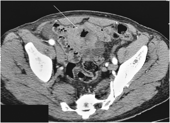
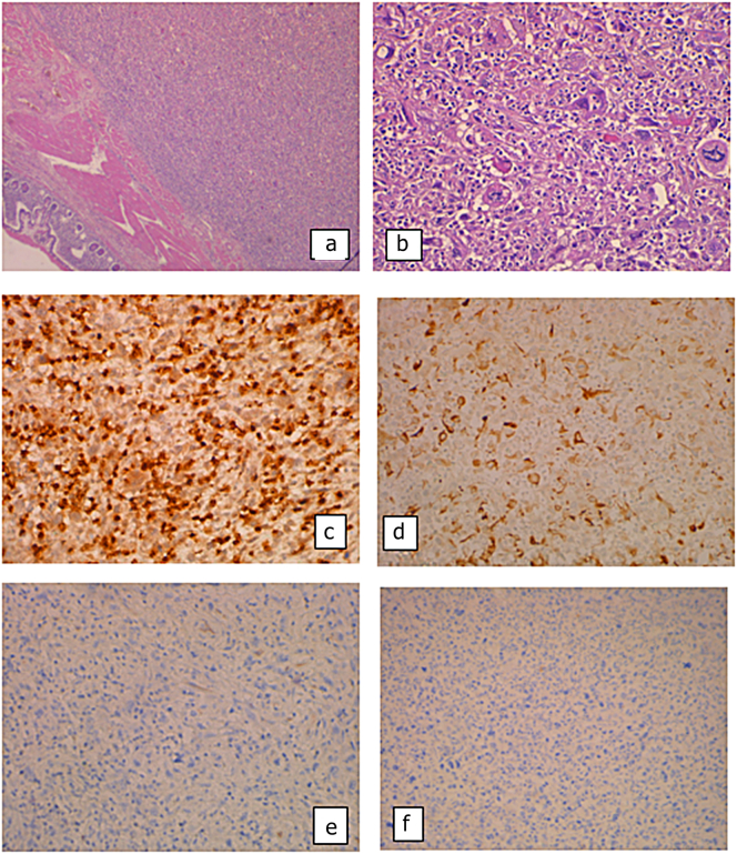
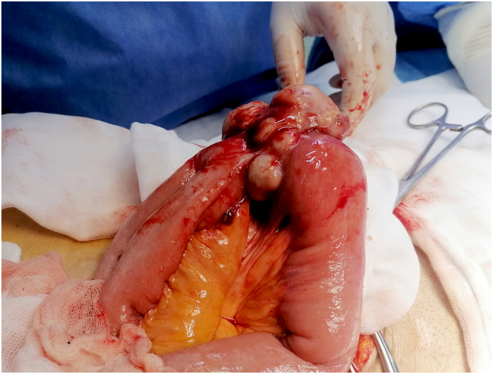
A 65-year-old patient, with no comorbidities, presented with chronic and paroxysmal abdominal pain. Upper endoscopy and colonoscopy showed no abnormalities. Thoracoabdominal computed tomography (CT) revealed an ileal lobulated, heterogeneously enhancing solid mass measuring 6 cm. Laparotomy was performed. Findings showed a lobulated ileal mass. We made an enlarged ileal resection with end-to-end anastomosis. The postoperative course was uneventful. Histology and IHC stains concluded into ileal LMS. No relapse of the disease was noted during the 4-month follow-up.
Ileal LMS is a rare tumor originating from the smooth muscle cells within the muscularis mucosa or muscularis propria. CT colonography (CTC) and magnetic resonance enterography (MRE) represent good options to aid the diagnosis. Histologically, LMS often has a comparable morphological appearance to GISTs. IHC is essential to differentiate those tumors. Surgery is the only curative treatment. The prognosis is poor knowing that those tumors are discovered at advanced stages.
Ileal LMS is a rare tumor originating from the smooth muscle cells. It has a comparable morphological appearance to GISTs. Immunohistochemistry is essential to confirm the diagnosis. Surgery is the only curative treatment. The prognosis is poor.
小肠恶性肿瘤较为罕见。空肠、回肠和十二指肠是肠道平滑肌肉瘤(LMS)最常见的发病部位。在此,我们报告一例65岁回肠LMS患者经手术切除成功治疗的病例。
一名65岁无合并症患者,出现慢性阵发性腹痛。上消化道内镜检查和结肠镜检查均未发现异常。胸腹部计算机断层扫描(CT)显示回肠有一个分叶状、强化不均匀的实性肿块,大小为6厘米。遂行剖腹手术。术中发现一个分叶状回肠肿块。我们进行了扩大的回肠切除术并端端吻合。术后过程顺利。组织学和免疫组化染色结果确诊为回肠LMS。在4个月的随访期间未发现疾病复发。
回肠LMS是一种罕见的肿瘤,起源于黏膜肌层或固有肌层内的平滑肌细胞。CT结肠成像(CTC)和磁共振小肠造影(MRE)是辅助诊断的良好选择。在组织学上,LMS通常与胃肠道间质瘤(GIST)有相似的形态学表现。免疫组化对于鉴别这些肿瘤至关重要。手术是唯一的治愈性治疗方法。鉴于这些肿瘤在晚期才被发现,其预后较差。
回肠LMS是一种起源于平滑肌细胞的罕见肿瘤。它与GIST有相似的形态学表现。免疫组化对于确诊至关重要。手术是唯一的治愈性治疗方法。预后较差。