Anhui University of Traditional Chinese Medicine Affiliated Chuzhou Hospital of Integrated Chinese and Western Medicine, Chuzhou, China.
Southeast University Affiliated Zhongda Hospital, Nanjing, China.
Front Endocrinol (Lausanne). 2022 Jul 14;13:918212. doi: 10.3389/fendo.2022.918212. eCollection 2022.
The decline in the quantity and quality of oocytes due to ovarian ageing in women is now a significant threat to reproductive health today as the concept of delayed fertility becomes widespread. However, the molecular mechanisms of natural ovarian ageing have not been fully elucidated.
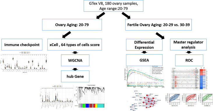
Here, we used transcriptomic data from 180 normal ovarian tissues from GTEx V8 to analyze the expression profile of ovarian tissues from women with age segments of 20-29 (22 individuals), 30-39 (14 individuals), 40-49 (37 individuals), 50-59 (61 individuals), 60-69 (42 individuals), and 70-79 (4 individuals), respectively. XCELL was used to assess the infiltration score of 64 cell types of the ovary. WGCNA was used to characterize the co-expression network during the natural aging of the ovary. ClusterprofileR was used for functional enrichment analysis of co-expression modules. MsViper was used for master regulator analysis.
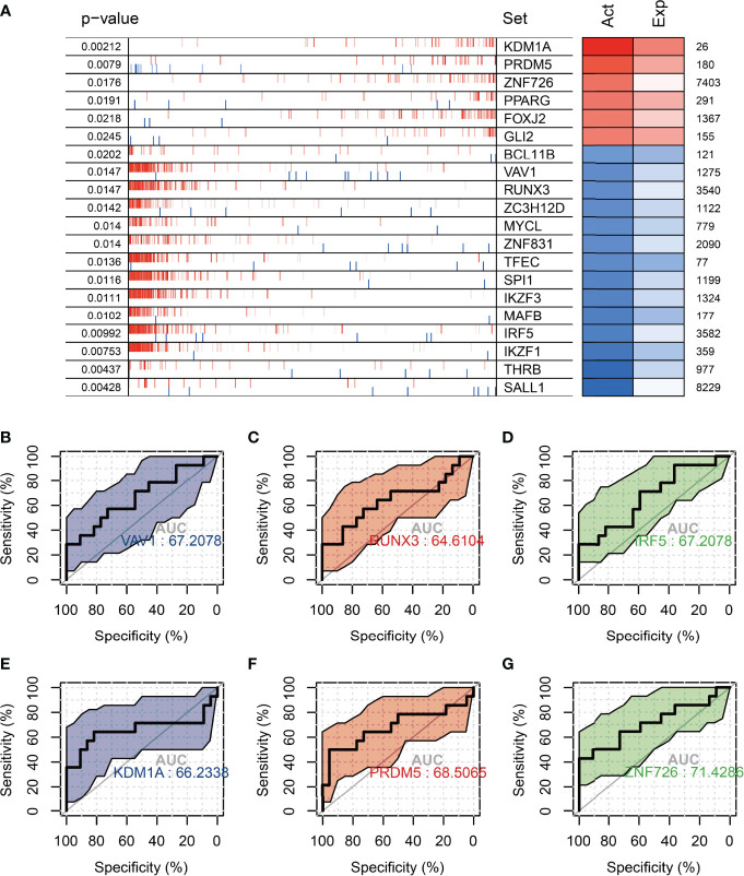
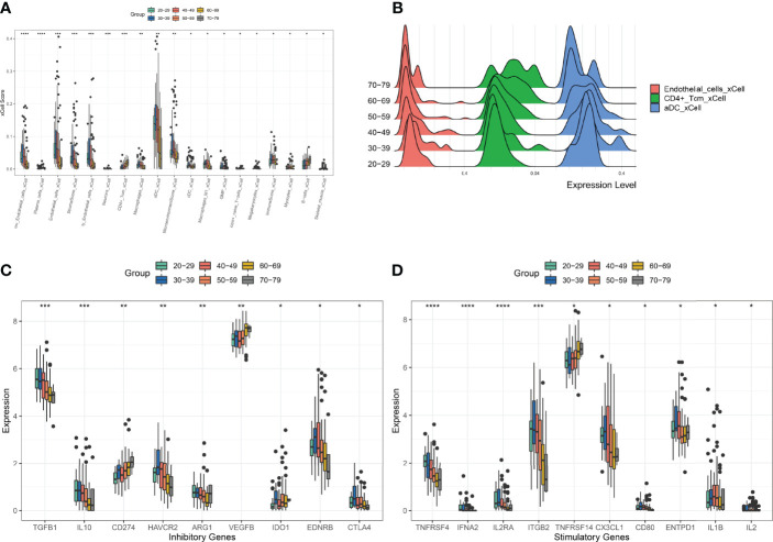
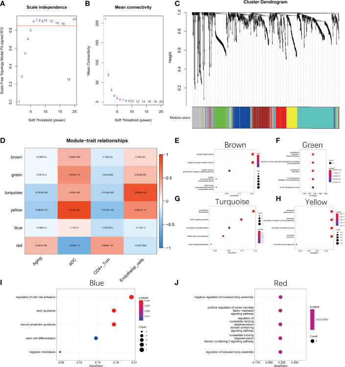
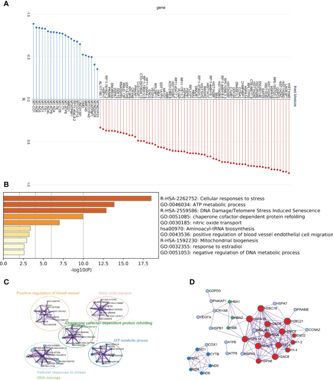
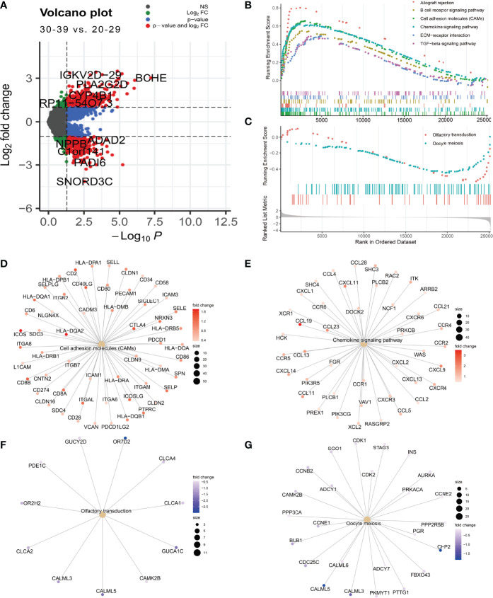
The infiltration score of endothelial cells and activated antigen-presenting cells during natural ovarian ageing increased significantly at ages 30-39, 40-49, and then decreased, whereas CD4+ Tcm increased with age. WGCNA identified six co-expression modules from ovarian tissue transcriptomic data species. The red module was significantly and positively correlated with senescence and CD4+ Tcm, and the turquoise module was significantly and positively correlated with Endothelial Cells. We further explored ovarian tissue for women aged 20-29 and 30-39 years. The GSEA results showed that the Chemokine signaling pathway was significantly activated in the 30-39-year-old group, while Oocyte meiosis was significantly inhibited. Finally, the results of msviper found that transcription factors such as KDM1A, PRDM5, ZNF726, PPARG, FOXJ2, and GLI2 were mainly activated in the 20-29 years group, while VAV1, RUNX3, ZC3H12D, MYCL, and IRF5 were mainly activated in the 30-39 years group and that these transcription factor activities were diagnostic of natural ovarian ageing (AUC: 0.65-0.71).
Natural ageing of the ovary is significantly correlated with immune cell infiltration and activation of inflammation-related signaling pathways, with inflammation levels reaching a maximum during early ovarian ageing (30-39, 40-49) and then gradually decreasing after that. These studies provide a research basis for exploring the mechanisms of natural ovarian ageing.
由于女性卵巢衰老导致的卵子数量和质量下降,如今对生殖健康构成了重大威胁,因为延迟生育的观念已经普及。然而,自然卵巢衰老的分子机制尚未完全阐明。
在这里,我们使用来自 GTEx V8 的 180 个正常卵巢组织的转录组数据,分析年龄在 20-29 岁(22 人)、30-39 岁(14 人)、40-49 岁(37 人)、50-59 岁(61 人)、60-69 岁(42 人)和 70-79 岁(4 人)的女性卵巢组织的表达谱。使用 XCELL 评估卵巢 64 种细胞类型的浸润评分。使用 WGCNA 对卵巢自然衰老过程中的共表达网络进行特征描述。使用 ClusterprofileR 对共表达模块进行功能富集分析。使用 MsViper 进行主调控因子分析。
在 30-39 岁、40-49 岁时,自然卵巢衰老过程中内皮细胞和激活抗原呈递细胞的浸润评分显著增加,然后下降,而 CD4+ Tcm 则随着年龄的增长而增加。WGCNA 从卵巢组织转录组数据中鉴定出六个共表达模块。红色模块与衰老和 CD4+ Tcm 呈显著正相关,而蓝色模块与内皮细胞呈显著正相关。我们进一步研究了 20-29 岁和 30-39 岁女性的卵巢组织。GSEA 结果表明,30-39 岁组趋化因子信号通路显著激活,而卵母细胞减数分裂显著受到抑制。最后,msviper 的结果发现,KDM1A、PRDM5、ZNF726、PPARG、FOXJ2 和 GLI2 等转录因子在 20-29 岁组主要激活,而 VAV1、RUNX3、ZC3H12D、MYCL 和 IRF5 等转录因子在 30-39 岁组主要激活,这些转录因子的活性可诊断自然卵巢衰老(AUC:0.65-0.71)。
卵巢的自然衰老与免疫细胞浸润和炎症相关信号通路的激活显著相关,炎症水平在早期卵巢衰老(30-39、40-49 岁)时达到最高,然后逐渐下降。这些研究为探索自然卵巢衰老的机制提供了研究基础。