Randall Centre for Cell & Molecular Biophysics, King's College London, London, UK.
Centre for Human & Applied Physiological Sciences, King's College London, London, UK.
J Physiol. 2022 Sep;600(17):3983-4000. doi: 10.1113/JP283048. Epub 2022 Aug 14.
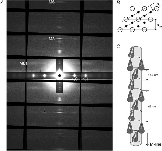
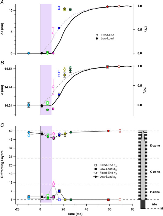
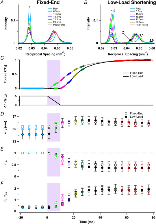
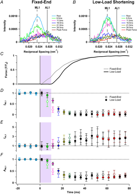
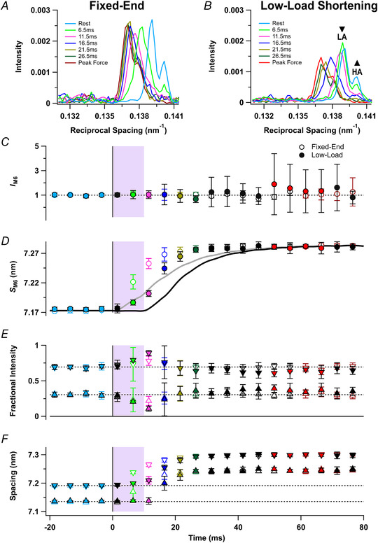
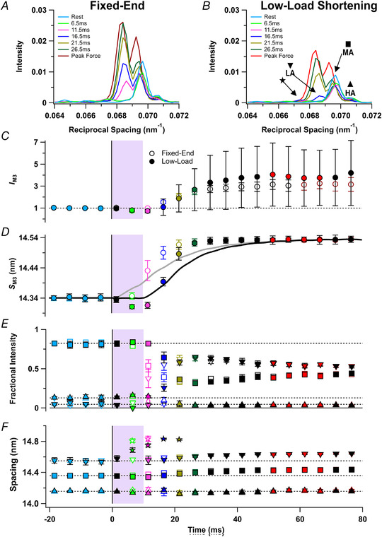
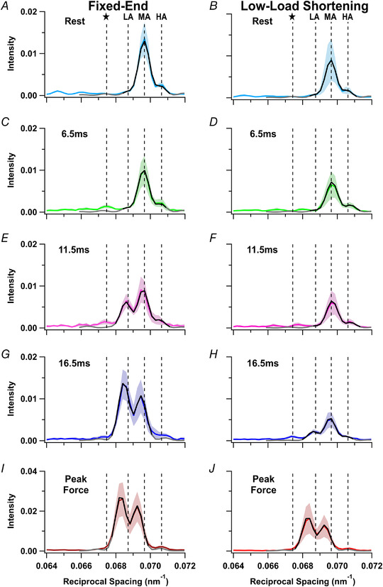
Myosin motors in resting muscle are inactivated by folding against the backbone of the myosin filament in an ordered helical array and must be released from that conformation to engage in force generation. Time-resolved X-ray diffraction from single fibres of amphibian muscle showed that myosin filament activation could be inhibited by imposing unloaded shortening at the start of stimulation, suggesting that filaments were activated by mechanical stress. Here we improved the signal-to-noise ratio of that approach using whole extensor digitorum longus muscles of the mouse contracting tetanically at 28°C. Changes in X-ray signals associated with myosin filament activation, including the decrease in the first-order myosin layer line associated with the helical motor array, increase in the spacing of a myosin-based reflection associated with packing of myosin tails in the filament backbone, and increase in the ratio of the 1,1 and 1,0 equatorial reflections associated with movement of motors away from the backbone, were delayed by imposing 10-ms unloaded shortening at the start of stimulation. These results show that myosin filaments are predominantly activated by filament stress, as in amphibian muscle. However, a small component of filament activation at zero load was detected, implying an independent mechanism of partial filament activation. X-ray interference measurements indicated a switch-like change in myosin motor conformation at the start of force development, accompanied by transient disordering of motors in the regions of the myosin filament near its midpoint, suggesting that filament zonal dynamics also play a role in its activation. KEY POINTS: Activation of myosin filaments in extensor digitorum longus muscles of the mouse is delayed by imposing rapid shortening from the start of stimulation. Stress is the major mechanism of myosin filament activation in these muscles, but there is a small component of filament activation during electrical stimulation at zero stress. Myosin motors switch rapidly from the folded inhibited conformation to the actin-attached force-generating conformation early in force development.
在休息状态下的肌肉中,肌球蛋白分子马达通过折叠在肌球蛋白丝的骨架上形成有序的螺旋阵列而失活,必须从该构象中释放出来才能进行力的产生。来自两栖动物肌肉的单纤维的时间分辨 X 射线衍射表明,肌球蛋白丝的激活可以通过在刺激开始时施加空载缩短来抑制,这表明纤维的激活是通过机械应力实现的。在这里,我们使用 28°C 下收缩的小鼠伸趾长肌的整个伸肌,改进了该方法的信噪比。与肌球蛋白丝激活相关的 X 射线信号变化,包括与螺旋马达阵列相关的第一阶肌球蛋白层线的减少、与肌球蛋白尾部在丝骨架中的包装相关的基于肌球蛋白的反射的间距增加,以及与马达远离骨架运动相关的 1,1 和 1,0 赤道反射的比值增加,在刺激开始时施加 10ms 的空载缩短会被延迟。这些结果表明,肌球蛋白丝主要通过纤维应力激活,就像在两栖动物肌肉中一样。然而,在零负载下检测到肌球蛋白丝激活的一小部分,这意味着存在部分肌球蛋白丝激活的独立机制。X 射线干涉测量表明,在力发展开始时,肌球蛋白马达构象发生类似开关的变化,同时在肌球蛋白丝中部附近的马达区域出现短暂的无序,这表明纤维区域动力学也在其激活中起作用。关键点:在刺激开始时施加快速缩短会延迟小鼠伸趾长肌中肌球蛋白丝的激活。在这些肌肉中,应力是肌球蛋白丝激活的主要机制,但在零应力下进行电刺激时,肌球蛋白丝会有一小部分激活。在力发展的早期,肌球蛋白马达迅速从折叠的抑制构象切换到与肌动蛋白结合的产生力的构象。