Akhter Nadeem, Kochumon Shihab, Hasan Amal, Wilson Ajit, Nizam Rasheeba, Al Madhoun Ashraf, Al-Rashed Fatema, Arefanian Hossein, Alzaid Fawaz, Sindhu Sardar, Al-Mulla Fahd, Ahmad Rasheed
Immunology & Microbiology Department, Dasman Diabetes Institute, Kuwait City, Kuwait.
Genetics & Bioinformatics, Dasman Diabetes Institute, Kuwait City, Kuwait.
J Inflamm Res. 2022 Jul 27;15:4291-4302. doi: 10.2147/JIR.S368352. eCollection 2022.
Overexpression of CCL2 (MCP-1) has been implicated in pathogenesis of metabolic conditions, such as obesity and T2D. However, the mechanisms leading to increased CCL2 expression in obesity are not fully understood. Since both IFN-γ and LPS levels are found to be elevated in obesity and shown to be involved in the regulation of metabolic inflammation and insulin resistance, we investigated whether these two agents could synergistically trigger the expression of CCL2 in obesity.
Monocytes (Human monocytic THP-1 cells) were stimulated with IFN-γ and LPS. CCL2 gene expression was determined by real-time RT-PCR. CCL2 protein was determined by ELISA. Signaling pathways were identified by using epigenetic inhibitors and STAT1 siRNA. Acetylation of H3K27 was analyzed by Western blotting. The acetylation level of histone H3K27 in the transcriptional initiation region of CCL2 gene was determined by ChIP-qPCR.
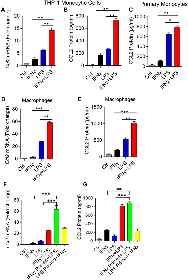
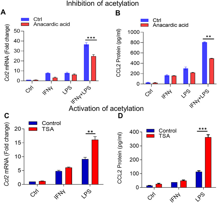
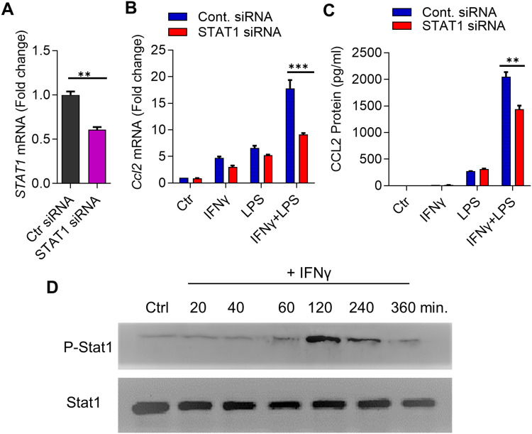
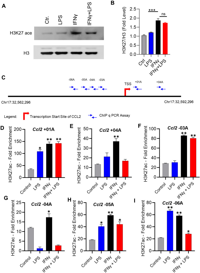
Our results show that the co-incubation of THP-1 monocytes with IFN-γ and LPS significantly enhanced the expression of CCL2, compared to treatment with IFN-γ or LPS alone. Similar results were obtained using primary monocytes and macrophages. Interestingly, IFN-γ priming was found to be more effective than LPS priming in inducing synergistic expression of CCL2. Moreover, STAT1 deficiency significantly suppressed this synergy for CCL2 expression. Mechanistically, we showed that IFN-γ priming induced acetylation of lysine 27 on histone 3 (H3K27ac) in THP-1 cells. Chromatin immunoprecipitation (ChIP) assay followed by qRT-PCR revealed increased H3K27ac at the CCL2 promoter proximal region, resulting in stabilized gene expression. Furthermore, inhibition of histone acetylation with anacardic acid suppressed this synergistic response, whereas trichostatin A (TSA) could substitute IFN-γ in this synergy.
Our findings suggest that IFN-γ, in combination with LPS, has the potential to augment inflammation via the H3K27ac-mediated induction of CCL2 in monocytic cells in the setting of obesity.
CCL2(单核细胞趋化蛋白-1)的过表达与肥胖和2型糖尿病等代谢性疾病的发病机制有关。然而,肥胖中导致CCL2表达增加的机制尚未完全明确。由于在肥胖中发现干扰素-γ(IFN-γ)和脂多糖(LPS)水平均升高,且二者均参与代谢性炎症和胰岛素抵抗的调节,因此我们研究了这两种因子是否能协同触发肥胖中CCL2的表达。
用IFN-γ和LPS刺激单核细胞(人单核细胞THP-1细胞)。通过实时逆转录聚合酶链反应(RT-PCR)测定CCL2基因表达。通过酶联免疫吸附测定(ELISA)测定CCL2蛋白。使用表观遗传抑制剂和信号转导和转录激活因子1(STAT1)小干扰RNA(siRNA)鉴定信号通路。通过蛋白质免疫印迹法分析组蛋白H3赖氨酸27(H3K27)的乙酰化。通过染色质免疫沉淀定量聚合酶链反应(ChIP-qPCR)测定CCL2基因转录起始区域的组蛋白H3K27乙酰化水平。
我们的结果表明,与单独用IFN-γ或LPS处理相比,THP-1单核细胞与IFN-γ和LPS共同孵育可显著增强CCL2的表达。使用原代单核细胞和巨噬细胞也获得了类似结果。有趣的是,在诱导CCL2协同表达方面,IFN-γ预处理比LPS预处理更有效。此外,STAT1缺陷显著抑制了CCL2表达的这种协同作用。机制上,我们发现IFN-γ预处理可诱导THP-1细胞中组蛋白3赖氨酸27(H3K27ac)的乙酰化。染色质免疫沉淀(ChIP)分析后进行定量逆转录聚合酶链反应(qRT-PCR)显示CCL2启动子近端区域的H3K27ac增加,从而导致基因表达稳定。此外,用漆树酸抑制组蛋白乙酰化可抑制这种协同反应,而曲古抑菌素A(TSA)可在这种协同作用中替代IFN-γ。
我们的研究结果表明,在肥胖情况下,IFN-γ与LPS联合有潜力通过H3K27ac介导的CCL2诱导增强单核细胞中的炎症反应。