Akhter Nadeem, Hasan Amal, Shenouda Steve, Wilson Ajit, Kochumon Shihab, Ali Shamsha, Tuomilehto Jaakko, Sindhu Sardar, Ahmad Rasheed
Immunology Unit, Dasman Diabetes Institute (DDI), Al-Soor Street, P.O. Box 1180, 15462 Dasman, Kuwait.
J Diabetes Metab Disord. 2018 Apr 16;17(1):77-84. doi: 10.1007/s40200-018-0341-y. eCollection 2018 Jun.
Obese human and mice were reported to have higher circularity endotoxin (LPS) levels as compared to their lean counter parts. The current study was aimed to reveal the molecular mechanisms underlying the LPS mediated induction of CCL2 in human monocytes/macrophages.
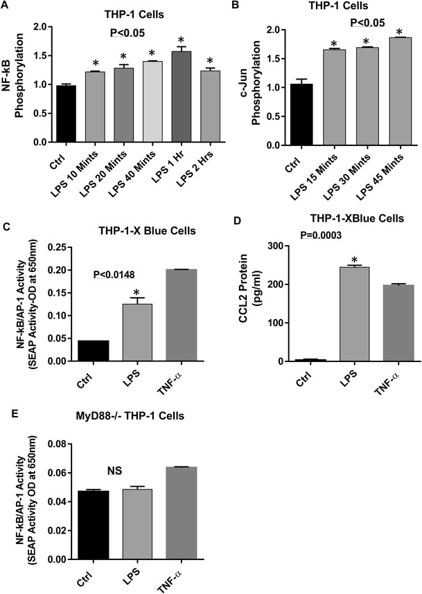
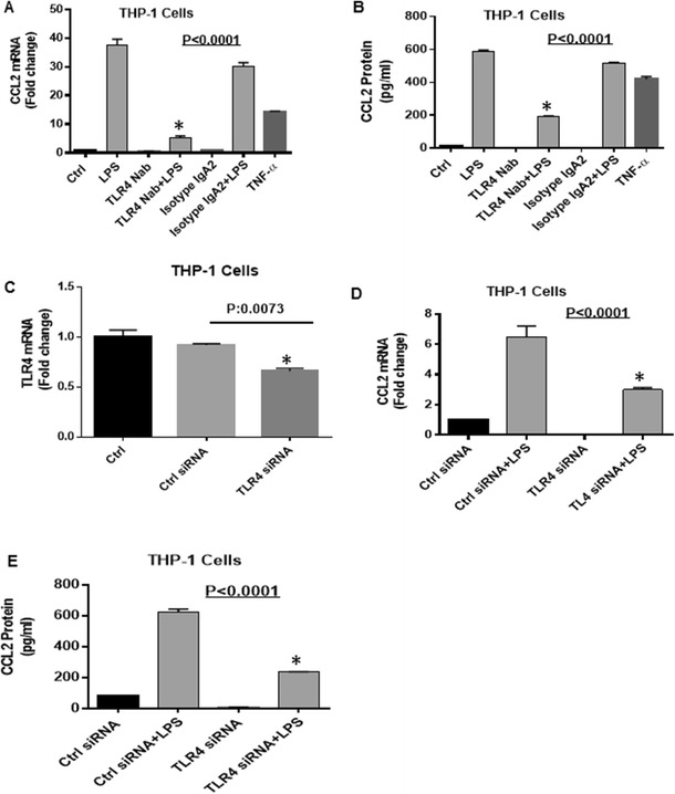
Human monocytic cell line THP-1, THP-1 cells derived macrophages and primary macrophages were treated with LPS and TNF-α (positive control). CCL2 expression was determined with real-time RT-PCR and ELISA. THP-1-XBlue™ cells, THP-1-XBlue™-defMyD cells, TLR4 neutralization antibody, TLR4 siRNA and inhibitors for NF-kB and MAPK were used to study the signaling pathways. Phosphorylation of NF-kB and c-Jun was analyzed by ELISA.
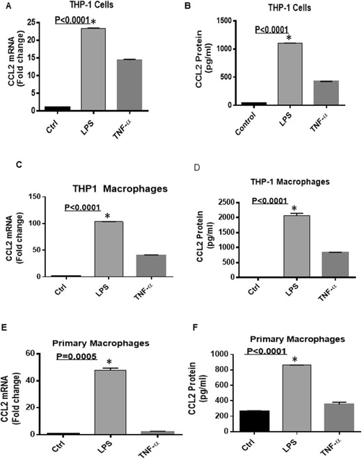
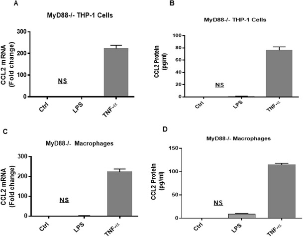
LPS upregulates CCL2 expression at both mRNA (THP-1: 23.40 ± .071 Fold, 0.0001; THP-1-derived macrophages: 103 ± 0.56 Fold, 0.0001; Primary macrophages: 48 ± 1.41 Fold, < 0.0005) and protein (THP1 monocytes:1048 ± 5.67 pg/ml, < 0.0001; THP-1-derived macrophages; 2014 ± 2.12, = 0.0001; Primary macrophages: 859.5 ± 3.54, < 0.0001) levels in human monocytic cells/macrophages. Neutralization of TLR4 blocked LPS-induced CCL-2 secretion ( < 0.0001). Silencing of TLR4 by siRNA also significantly reduced LPS-induced CCL-2 production. Furthermore, MyD88-Knockout cells treated with LPS did not produce CCL-2. NF-kB and c-Jun phosphorylation was noted in LPS treated cells.
Overall, our data reveal that LPS induces CCL-2 in monocytes/macrophages via TLR4/MyD88 signaling which leads to the activation of NF-kB/AP-1 transcription factors.
据报道,肥胖的人类和小鼠体内的循环内毒素(LPS)水平高于体型偏瘦的同类个体。本研究旨在揭示LPS介导人单核细胞/巨噬细胞中CCL2诱导的分子机制。
用LPS和TNF-α(阳性对照)处理人单核细胞系THP-1、THP-1来源的巨噬细胞和原代巨噬细胞。用实时RT-PCR和ELISA检测CCL2表达。使用THP-1-XBlue™细胞、THP-1-XBlue™-defMyD细胞、TLR4中和抗体、TLR4 siRNA以及NF-κB和MAPK抑制剂来研究信号通路。通过ELISA分析NF-κB和c-Jun的磷酸化。
LPS在mRNA水平(THP-1:23.40±0.071倍,P<0.0001;THP-1来源的巨噬细胞:103±0.56倍,P<0.0001;原代巨噬细胞:48±1.41倍,P<0.0005)和蛋白水平(THP1单核细胞:1048±5.67 pg/ml,P<0.0001;THP-1来源的巨噬细胞:2014±2.12,P=0.0001;原代巨噬细胞:859.5±3.54,P<0.0001)上均上调人单核细胞/巨噬细胞中CCL2的表达。TLR4的中和阻断了LPS诱导的CCL-2分泌(P<0.0001)。用siRNA沉默TLR4也显著降低了LPS诱导的CCL-2产生。此外,用LPS处理的MyD88基因敲除细胞不产生CCL-2。在LPS处理的细胞中观察到NF-κB和c-Jun的磷酸化。
总体而言,我们的数据表明,LPS通过TLR4/MyD88信号通路在单核细胞/巨噬细胞中诱导CCL-2,这导致NF-κB/AP-1转录因子的激活。