Hung George, Ashvetiya Tamara, Leszczynska Aleksandra, Yang Wanjun, Hwang Chao-Wei, Gerstenblith Gary, Barth Andreas S, Johnston Peter V
Department of Medicine, Division of Cardiology, Johns Hopkins University School of Medicine, Baltimore, MD, USA.
Department of Medicine, Section of Cardiovascular Medicine, Yale University School of Medicine, New Haven, CT, USA.
NPJ Aging. 2022 Jul 18;8(1):10. doi: 10.1038/s41514-022-00091-0.
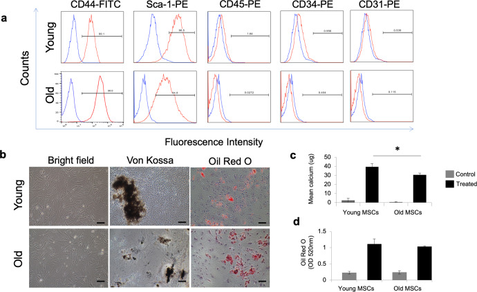
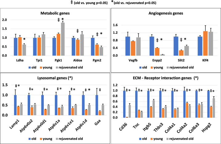
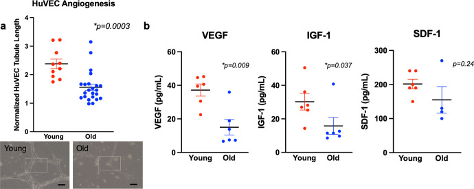
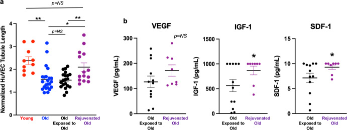
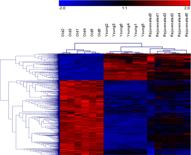
Age-related differences in stem-cell potency contribute to variable outcomes in clinical stem cell trials. To help understand the effect of age on stem cell potency, bone marrow-derived mesenchymal stem cells (MSCs) were isolated from young (6 weeks) and old (18-24 months) mice. HUVEC tubule formation (TF) induced by the old and young MSCs and ELISA of conditioned media were compared to one another, and to old MSCs after 7 d in indirect co-culture with young MSCs. Old MSCs induced less TF than did young (1.56 ± 0.11 vs 2.38 ± 0.17, p = 0.0003) and released lower amounts of VEGF (p = 0.009) and IGF1 (p = 0.037). After 7 d in co-culture with young MSCs, TF by the old MSCs significantly improved (to 2.09 ± 0.18 from 1.56 ± 0.11; p = 0.013), and was no longer different compared to TF from young MSCs (2.09 ± 0.18 vs 2.38 ± 0.17; p = 0.27). RNA seq of old MSCs, young MSCs, and old MSCs following co-culture with young MSCs revealed that the age-related differences were broadly modified by co-culture, with the most significant changes associated with lysosomal pathways. These results indicate that the age-associated decreased paracrine-mediated effects of old MSCs are improved following indirect co-culture with young MSC. The observed effect is associated with broad transcriptional modification, suggesting potential targets to both assess and improve the therapeutic potency of stem cells from older patients.
干细胞潜能的年龄相关差异导致临床干细胞试验结果各异。为了帮助理解年龄对干细胞潜能的影响,从年轻(6周龄)和年老(18 - 24月龄)小鼠中分离出骨髓间充质干细胞(MSC)。比较了年轻和年老MSC诱导的人脐静脉内皮细胞(HUVEC)管腔形成(TF)以及条件培养基的酶联免疫吸附测定(ELISA),并将年老MSC与年轻MSC间接共培养7天后的情况进行比较。年老MSC诱导的TF少于年轻MSC(1.56±0.11对2.38±0.17,p = 0.0003),并且释放的血管内皮生长因子(VEGF)(p = 0.009)和胰岛素样生长因子1(IGF1)(p = 0.037)量更低。与年轻MSC共培养7天后,年老MSC的TF显著改善(从1.56±0.11提高到2.09±0.18;p = 0.013),与年轻MSC的TF相比不再有差异(2.09±0.18对2.38±0.17;p = 0.27)。对年老MSC、年轻MSC以及与年轻MSC共培养后的年老MSC进行RNA测序,结果显示年龄相关差异通过共培养得到广泛改变,最显著的变化与溶酶体途径相关。这些结果表明,年老MSC与年龄相关的旁分泌介导效应在与年轻MSC间接共培养后得到改善。观察到的效应与广泛的转录修饰相关,提示了评估和提高老年患者干细胞治疗潜能的潜在靶点。