Prenatal Medicine Unit, Obstetrics and Gynecology Unit, Department of Medical and Surgical Sciences for Mother, Child and Adult, University of Modena and Reggio Emilia, Modena, Italy.
Department of Biomedical, Metabolic and Neural Sciences, International Doctorate School in Clinical and Experimental Medicine, University of Modena and Reggio Emilia, Modena, Italy.
Int J Gynaecol Obstet. 2023 Mar;160(3):856-863. doi: 10.1002/ijgo.14383. Epub 2022 Aug 17.
To study how adenomyosis changes during pregnancy and to possibly correlate these changes to maternal and fetal outcomes.
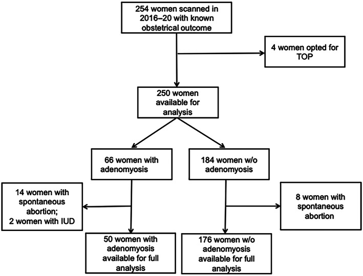
Retrospective exploratory cohort study including 254 women with a pre-conceptional/first-trimester scan to document adenomyosis and known obstetric outcome. If visible, adenomyosis signs were documented in each trimester and postpartum. Mann-Whitney U tests or χ tests were used for continuous and categorical variables, respectively.
A globular uterus was reported in 79% (n = 52) of women with adenomyosis in the first trimester, in 38% (n = 20) and 2% (n = 1) of women in the second and third trimesters, respectively, and postpartum in 77% (n = 34) of women. Asymmetrical thickening (n = 20, 30%) and cysts (n = 15, 23%) were only visible in 1st trimester. Adenomyosis was associated with miscarriage (odds ratio [OR] 5.9, 95% confidence interval [CI] 2.4-14.9, P < 0.001) also in normal conception only (OR 5.1, 95% CI 1.8-14.2, P = 0.002) or adjusting for maternal age (adjusted OR 5.9, 95% CI 2.3-15.2, P < 0.001). Gestational age at delivery was lower in adenomyosis (P = 0.004); the cesarean section rate was higher than in controls (OR 2.5, 95% CI 1.3-4.8, P = 0.007) also adjusting for age (adjusted OR 2.07, 95% CI 1.06-4.08, P = 0.035).
Signs of adenomyosis were visible but progressively disappeared in pregnancy; adenomyosis was associated with an increased risk of early miscarriage. Prospective studies are needed to confirm our results.
研究子宫腺肌病在妊娠期间的变化,并可能将这些变化与母婴结局相关联。
本研究为回顾性探索性队列研究,纳入了 254 名在受孕前/孕早期接受扫描以记录子宫腺肌病和已知产科结局的女性。如果可见,在每个孕早期、孕中期和孕晚期以及产后记录子宫腺肌病的征象。连续变量和分类变量分别采用曼-惠特尼 U 检验或卡方检验。
在孕早期,79%(n=52)的子宫腺肌病女性报告子宫呈球形,38%(n=20)和 2%(n=1)的女性在孕中期和孕晚期以及产后报告子宫呈球形,分别为 77%(n=34)。不对称性增厚(n=20,30%)和囊肿(n=15,23%)仅在孕早期可见。子宫腺肌病与流产有关(比值比[OR] 5.9,95%置信区间[CI] 2.4-14.9,P<0.001),仅在正常妊娠中(OR 5.1,95% CI 1.8-14.2,P=0.002)或调整了产妇年龄后(调整后 OR 5.9,95% CI 2.3-15.2,P<0.001)。子宫腺肌病患者的分娩孕周较低(P=0.004);剖宫产率高于对照组(OR 2.5,95% CI 1.3-4.8,P=0.007),调整年龄后也更高(调整后 OR 2.07,95% CI 1.06-4.08,P=0.035)。
妊娠期间可见子宫腺肌病的征象,但逐渐消失;子宫腺肌病与早期流产的风险增加相关。需要前瞻性研究来证实我们的结果。