Department of General, Visceral, Transplant, Vascular and Pediatric Surgery, University Hospital Würzburg, Oberduerrbacherstrasse 6, 97080, Würzburg, Germany.
Department of Internal Medicine I, Division of Endocrinology and Diabetes, University Hospital Würzburg, Oberduerrbacherstrasse 6, 97080, Würzburg, Germany.
Langenbecks Arch Surg. 2022 Dec;407(8):3661-3669. doi: 10.1007/s00423-022-02648-9. Epub 2022 Aug 9.
A successful focused surgical approach in primary hyperparathyroidism (pHPT) relies on accurate preoperative localization of the parathyroid adenoma (PA). Most often, ultrasound is followed by [Tc]-sestamibi scintigraphy, but the value of this approach is disputed. Here, we evaluated the diagnostic approach in patients with surgically treated pHPT in our center with the aim to further refine preoperative diagnostic procedures.
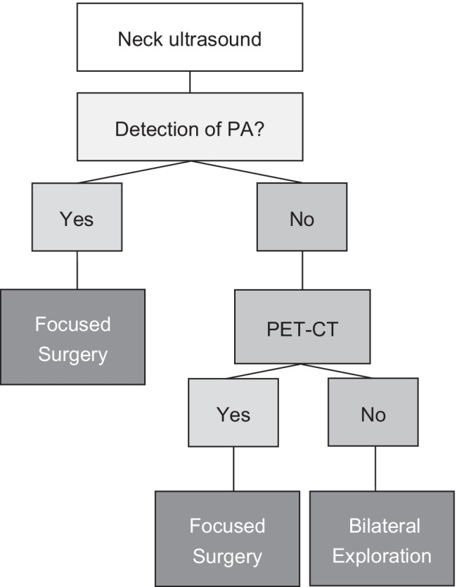
A single-center retrospective analysis of patients with pHPT from 01/2005 to 08/2021 was carried out followed by evaluation of the preoperative imaging modalities to localize PA. The localization of the PA had to be confirmed intraoperatively by the fresh frozen section and significant dropping of the intraoperative parathyroid hormone (PTH) levels.
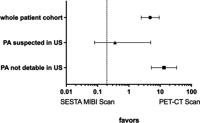
From 658 patients diagnosed with pHPT, 30 patients were excluded from the analysis because of surgery for recurrent or persistent disease. Median age of patients was 58.0 (13-93) years and 71% were female. Neck ultrasound was carried out in 91.7% and localized a PA in 76.6%. In 23.4% (135/576) of the patients, preoperative neck ultrasound did not detect a PA. In this group, [Tc]-sestamibi correctly identified PA in only 25.4% of patients. In contrast, in the same cohort, the use of [C]-methionine or [C]-choline PET resulted in the correct identification of PA in 79.4% of patients (OR 13.23; 95% CI 5.24-33.56).
[C]-Methionine or [C]-choline PET/CT are superior second-line imaging methods to select patients for a focused surgical approach when previous ultrasound failed to identify PA.
原发性甲状旁腺功能亢进症(pHPT)的成功聚焦手术方法依赖于甲状旁腺瘤(PA)的准确术前定位。通常,超声检查后会进行 [Tc]- sestamibi 闪烁扫描,但这种方法的价值存在争议。在这里,我们评估了我们中心接受手术治疗的 pHPT 患者的诊断方法,旨在进一步完善术前诊断程序。
对 2005 年 1 月至 2021 年 8 月在我们中心接受治疗的 pHPT 患者进行单中心回顾性分析,然后评估定位 PA 的术前影像学方法。PA 的定位必须通过新鲜冷冻切片在术中确认,并显著降低术中甲状旁腺激素(PTH)水平。
从 658 例 pHPT 患者中,有 30 例因手术治疗复发性或持续性疾病而被排除在分析之外。患者的中位年龄为 58.0(13-93)岁,71%为女性。91.7%的患者进行了颈部超声检查,76.6%的患者定位了 PA。在 23.4%(135/576)的患者中,术前颈部超声未检测到 PA。在这组患者中,[Tc]- sestamibi 仅正确识别出 25.4%的 PA。相比之下,在同一队列中,使用 [C]-蛋氨酸或 [C]-胆碱 PET 可使 79.4%的患者正确识别出 PA(OR 13.23;95%CI 5.24-33.56)。
当先前的超声检查未能识别出 PA 时,[C]-蛋氨酸或 [C]-胆碱 PET/CT 是选择聚焦手术方法的患者的优越二线成像方法。