• 文献检索
  • 文档翻译
  • 深度研究
  • 学术资讯
  • Suppr Zotero 插件Zotero 插件
  • 邀请有礼
  • 套餐&价格
  • 历史记录
应用&插件
Suppr Zotero 插件Zotero 插件浏览器插件Mac 客户端Windows 客户端微信小程序
定价
高级版会员购买积分包购买API积分包
服务
文献检索文档翻译深度研究API 文档MCP 服务
关于我们
关于 Suppr公司介绍联系我们用户协议隐私条款
关注我们

Suppr 超能文献

核心技术专利:CN118964589B侵权必究
粤ICP备2023148730 号-1Suppr @ 2026

文献检索

告别复杂PubMed语法,用中文像聊天一样搜索,搜遍4000万医学文献。AI智能推荐,让科研检索更轻松。

立即免费搜索

文件翻译

保留排版,准确专业,支持PDF/Word/PPT等文件格式,支持 12+语言互译。

免费翻译文档

深度研究

AI帮你快速写综述,25分钟生成高质量综述,智能提取关键信息,辅助科研写作。

立即免费体验

基于三维足底扫描仪和数字足迹分析扁平足骨骼特征。

Analysis of skeletal characteristics of flat feet using three-dimensional foot scanner and digital footprint.

机构信息

Graduate School of Engineering, Osaka City University, 3-3-138 Sugimoto, Sumiyoshi, Osaka, 558-8585, Japan.

Department of Clinical Engineering, Faculty of Human Care at Makuhari, Tohto University, Chiba, Japan.

出版信息

Biomed Eng Online. 2022 Aug 9;21(1):56. doi: 10.1186/s12938-022-01021-7.

DOI:10.1186/s12938-022-01021-7
PMID:35945533
原文链接:https://pmc.ncbi.nlm.nih.gov/articles/PMC9361570/
Abstract

BACKGROUND

Flat feet increase the risk of knee osteoarthritis and contribute to frailty, which may lead to worse life prognoses. The influence of the foot skeletal structure on flat feet is not yet entirely understood. Footprints are often used to evaluate feet. However, footprint-based measurements do not reflect the underlying structures of feet and are easily confounded by soft tissue. Three-dimensional evaluation of the foot shape can reveal the characteristics of flat feet. Therefore, foot shape evaluations have garnered increasing research interest. This study aimed to determine the correlation between the three-dimensional (3D) features of the foot and the measurement results of footprint and to predict the evaluation results of flat feet from the footprint based on the 3D features. Finally, the three-dimensional characteristics of flat feet, which cannot be revealed by footprint, were determined.

METHODS

A total of 403 individuals (40-89 years) participated in this study. The proposed system was developed to identify seven skeletal features that were expected to be associated with flat feet. The loads on the soles of the feet were measured in a static standing position and with a digital footprint device. Specifically, two footprint indices were calculated: the Chippaux-Smirak index (CSI) and the Staheli index (SI). In the analysis, comparisons between male and female measurement variables were performed using the Student's t test. The relationships between the 3D foot features and footprint index parameters were determined by employing the Pearson correlation coefficient. Multiple linear regression was utilized to identify 3D foot features that were strongly associated with the CSI and SI. Foot features identified as significant in the multivariate regression analysis were compared based on a one-way analysis of variance (ANOVA) with Tukey's post hoc test.

RESULTS

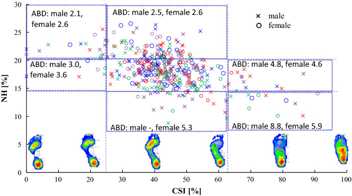
The CSI and SI were highly correlated with the instep height (IH) and navicular height (NH) of the 3D foot scanning system and were also derived from multiple regression analysis. In addition to the NH and IH, the indicators of the forefoot, transverse arch width, and transverse arch height were considered. In the flat foot group with CSI values above 62.7%, NH was 13.5% (p < 0.001) for males and 14.9% (p = 0.01) for females, and the axis of the bone distance was 5.3% (p = 0.05) for males and 4.9% (p = 0.10) for females. In particular, for CSI values above 62.7% and NH values below 13%, the axis of the bone distance was large and the foot skeleton was deformed.

CONCLUSIONS

Decreased navicular bone height could be evaluated with the 3D foot scanning system even when flat feet were not detected from the footprint. The results indicate that the use of quantitative indices for 3D foot measurements is important when evaluating the flattening of the foot. Trial registration number UMIN000037694. Name of the registry: University Hospital Medical Information Network Registry. Date of registration: August 15, 2019.

https://cdn.ncbi.nlm.nih.gov/pmc/blobs/6522/9361570/f2dc5b20be02/12938_2022_1021_Fig3_HTML.jpg
https://cdn.ncbi.nlm.nih.gov/pmc/blobs/6522/9361570/3064e90a70b3/12938_2022_1021_Fig1_HTML.jpg
https://cdn.ncbi.nlm.nih.gov/pmc/blobs/6522/9361570/92ae3e282fe7/12938_2022_1021_Fig2_HTML.jpg
https://cdn.ncbi.nlm.nih.gov/pmc/blobs/6522/9361570/f2dc5b20be02/12938_2022_1021_Fig3_HTML.jpg
https://cdn.ncbi.nlm.nih.gov/pmc/blobs/6522/9361570/3064e90a70b3/12938_2022_1021_Fig1_HTML.jpg
https://cdn.ncbi.nlm.nih.gov/pmc/blobs/6522/9361570/92ae3e282fe7/12938_2022_1021_Fig2_HTML.jpg
https://cdn.ncbi.nlm.nih.gov/pmc/blobs/6522/9361570/f2dc5b20be02/12938_2022_1021_Fig3_HTML.jpg
摘要

背景

扁平足会增加膝关节骨关节炎的风险,并导致虚弱,从而导致预后更差。足部骨骼结构对扁平足的影响尚不完全清楚。足印通常用于评估足部。然而,基于足印的测量并不能反映足部的潜在结构,并且很容易受到软组织的干扰。足部形状的三维评估可以揭示扁平足的特征。因此,对足部形状的评估越来越受到关注。本研究旨在确定足部的三维(3D)特征与足印测量结果之间的相关性,并基于 3D 特征预测基于足印的扁平足评估结果。最后,确定了足印无法揭示的扁平足的三维特征。

方法

共有 403 名(40-89 岁)个体参与了这项研究。该系统旨在识别七个预计与扁平足相关的骨骼特征。在静态站立位和数字足印设备上测量足底的负荷。具体来说,计算了两个足印指数:Chippaux-Smirak 指数(CSI)和 Staheli 指数(SI)。在分析中,使用学生 t 检验比较了男性和女性测量变量之间的差异。通过 Pearson 相关系数确定了 3D 足特征与足印指数参数之间的关系。采用多元线性回归确定与 CSI 和 SI 强烈相关的 3D 足特征。在多元回归分析中确定为显著的足特征,基于方差分析(ANOVA)与 Tukey 事后检验进行比较。

结果

CSI 和 SI 与 3D 足部扫描系统的足背高度(IH)和舟骨高度(NH)高度相关,也可通过多元回归分析得出。除了 NH 和 IH 之外,还考虑了前足、横弓宽度和横弓高度的指标。在 CSI 值大于 62.7%的扁平足组中,男性 NH 值为 13.5%(p<0.001),女性 NH 值为 14.9%(p=0.01),骨骼轴距离为 5.3%(p=0.05),女性为 4.9%(p=0.10)。特别是对于 CSI 值大于 62.7%且 NH 值低于 13%的情况,骨骼轴距离较大且足部骨骼变形。

结论

即使从足印中未检测到扁平足,也可以使用 3D 足部扫描系统评估舟骨高度降低。结果表明,在评估足部扁平时,使用 3D 足部测量的定量指标很重要。试验注册号 UMIN000037694。注册机构名称:大学医院医疗信息网注册处。注册日期:2019 年 8 月 15 日。

相似文献

1
Analysis of skeletal characteristics of flat feet using three-dimensional foot scanner and digital footprint.基于三维足底扫描仪和数字足迹分析扁平足骨骼特征。
Biomed Eng Online. 2022 Aug 9;21(1):56. doi: 10.1186/s12938-022-01021-7.
2
Variability between Clarke's angle and Chippaux-Smirak index for the diagnosis of flat feet.用于扁平足诊断的克拉克角与奇波-斯米拉指数之间的变异性。
Colomb Med (Cali). 2017 Mar 30;48(1):25-31.
3
Exploring flatfeet morphology in children aged 6-12 years: relationships with body mass and body height through footprints and three-dimensional measurements.探讨 6-12 岁儿童平足形态:通过足迹和三维测量与体重和身高的关系。
Eur J Pediatr. 2024 Apr;183(4):1901-1910. doi: 10.1007/s00431-024-05471-0. Epub 2024 Feb 9.
4
Describing the medial longitudinal arch using footprint indices and a clinical grading system.使用足迹指数和临床分级系统描述内侧纵弓。
Foot Ankle Int. 2007 Apr;28(4):456-62. doi: 10.3113/FAI.2007.0456.
5
The relationship between body mass index and footprint parameters in older people.老年人身体质量指数与足迹参数之间的关系。
Foot (Edinb). 2014 Dec;24(4):186-9. doi: 10.1016/j.foot.2014.08.009. Epub 2014 Aug 19.
6
Predictors of flatfoot in 11-12-year olds: a longitudinal cohort study.11-12 岁青少年扁平足的预测因素:一项纵向队列研究。
Biomed Eng Online. 2024 Aug 21;23(1):83. doi: 10.1186/s12938-024-01282-4.
7
Medial Longitudinal Arch: Accuracy, Reliability, and Correlation Between Navicular Drop Test and Footprint Parameters.内侧纵弓:舟骨下降试验与足迹参数之间的准确性、可靠性及相关性
J Manipulative Physiol Ther. 2018 Oct;41(8):672-679. doi: 10.1016/j.jmpt.2018.04.001. Epub 2018 Dec 17.
8
Would foot arch development in children characterize a body maturation process? A prospective longitudinal study.儿童足弓发育能否表征身体成熟过程?一项前瞻性纵向研究。
Biomed J. 2022 Oct;45(5):828-837. doi: 10.1016/j.bj.2021.10.012. Epub 2021 Nov 2.
9
Comparative assessment of flatfoot in preschool children.学龄前儿童扁平足的比较评估
Bol Med Hosp Infant Mex. 2020;77(6):312-319. doi: 10.24875/BMHIM.20000135.
10
Footprint analysis of flatfoot in preschool-aged children.学龄前儿童扁平足的足迹分析。
Eur J Pediatr. 2011 May;170(5):611-7. doi: 10.1007/s00431-010-1330-4. Epub 2010 Oct 23.

引用本文的文献

1
Biomechanics of transverse axis of medial longitudinal arch of children's foot based on 3D scanning.基于三维扫描的儿童足内侧纵弓横轴生物力学研究
Front Pediatr. 2023 Jul 10;11:1197439. doi: 10.3389/fped.2023.1197439. eCollection 2023.

本文引用的文献

1
Foot-surface-structure analysis using a smartphone-based 3D foot scanner.基于智能手机的 3D 足部扫描仪的足部结构分析。
Med Eng Phys. 2021 Sep;95:90-96. doi: 10.1016/j.medengphy.2021.08.001. Epub 2021 Aug 10.
2
Validity of a simple footprint assessment board for diagnosing the severity of flatfoot: a prospective cohort study.一种用于诊断扁平足严重程度的简易足迹评估板的效度:一项前瞻性队列研究。
BMC Musculoskelet Disord. 2021 Mar 18;22(1):285. doi: 10.1186/s12891-021-04154-3.
3
Development of a quantitative measurement system for three-dimensional analysis of foot morphology using a smartphone.
利用智能手机开发用于足部形态三维分析的定量测量系统。
Annu Int Conf IEEE Eng Med Biol Soc. 2019 Jul;2019:3171-3174. doi: 10.1109/EMBC.2019.8857209.
4
Association of quadriceps angle with plantar pressure distribution, navicular height and calcaneo-tibial angle.股四头肌角与足底压力分布、舟骨高度及跟骨-胫骨角的相关性
Acta Orthop Traumatol Turc. 2019 Mar;53(2):145-149. doi: 10.1016/j.aott.2018.12.008. Epub 2019 Jan 9.
5
Paediatric flexible flat foot: how are we measuring it and are we getting it right? A systematic review.小儿柔性扁平足:我们如何测量它,以及我们做得对吗?一项系统综述。
J Foot Ankle Res. 2018 May 30;11:21. doi: 10.1186/s13047-018-0264-3. eCollection 2018.
6
Reported selection criteria for adult acquired flatfoot deformity and posterior tibial tendon dysfunction: Are they one and the same? A systematic review.成人获得性平足畸形和胫后肌腱功能障碍的报告选择标准:它们是相同的吗?一项系统评价。
PLoS One. 2017 Dec 1;12(12):e0187201. doi: 10.1371/journal.pone.0187201. eCollection 2017.
7
Prevalence of Flexible Flat Foot in Adults: A Cross-sectional Study.成人柔性扁平足的患病率:一项横断面研究。
J Clin Diagn Res. 2017 Jun;11(6):AC17-AC20. doi: 10.7860/JCDR/2017/26566.10059. Epub 2017 Jun 1.
8
Association of bilateral flat feet with knee pain and disability in patients with knee osteoarthritis: A cross-sectional study.膝骨关节炎患者双侧扁平足与膝关节疼痛及功能障碍的关联:一项横断面研究。
J Orthop Res. 2017 Nov;35(11):2490-2498. doi: 10.1002/jor.23565. Epub 2017 Apr 24.
9
Flexible flatfoot.柔性扁平足
North Clin Istanb. 2014 Aug 3;1(1):57-64. doi: 10.14744/nci.2014.29292. eCollection 2014.
10
Knee and ankle biomechanics with lateral wedges with and without a custom arch support in those with medial knee osteoarthritis and flat feet.患有内侧膝关节骨关节炎和平足的人群使用带或不带定制足弓支撑的外侧楔形鞋垫时的膝关节和踝关节生物力学
J Orthop Res. 2016 Sep;34(9):1597-605. doi: 10.1002/jor.23174. Epub 2016 Feb 5.