Nagaraja Chandrashekhar T, Ramalingaiah Arvinda H, Arimappamagan Arivazhagan, Mitra Saikat, Shukla Dhaval, Srinivas Dwarakanath, Krishna Shankar S, Mahadevan Anita
Department of Pathology, Shimoga Institute of Medical Sciences, Shivammoga, Karnataka, India.
Departments of Neuroimaging and Interventional Radiology, National Institute of Mental Health and Neurosciences, Bengaluru, Karnataka, India.
J Neurosci Rural Pract. 2022 Aug 7;13(3):495-509. doi: 10.1055/s-0042-1750707. eCollection 2022 Jul.
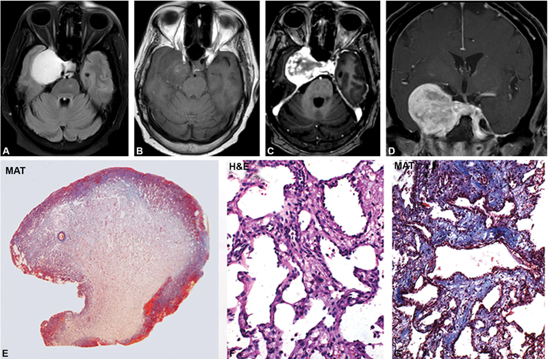
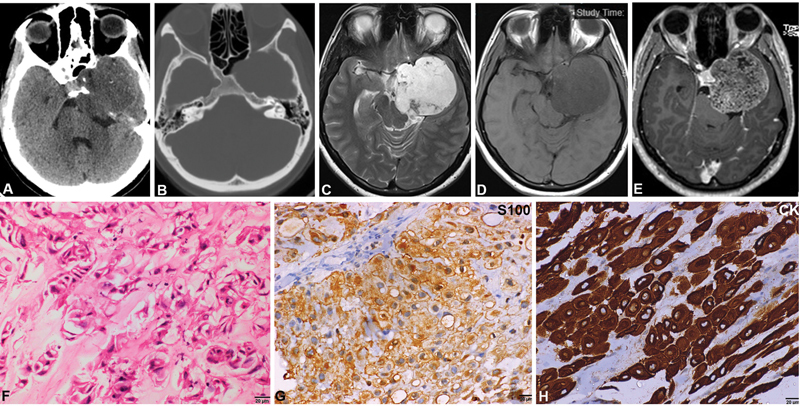
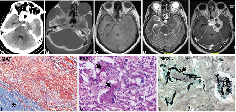
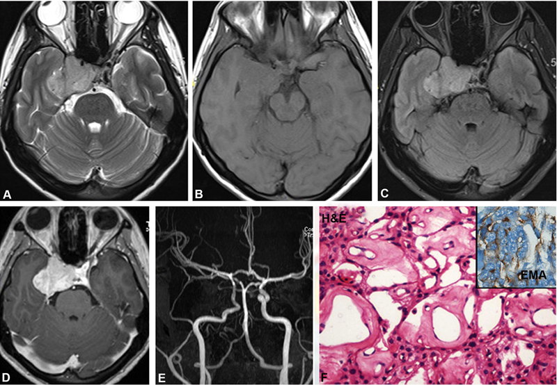
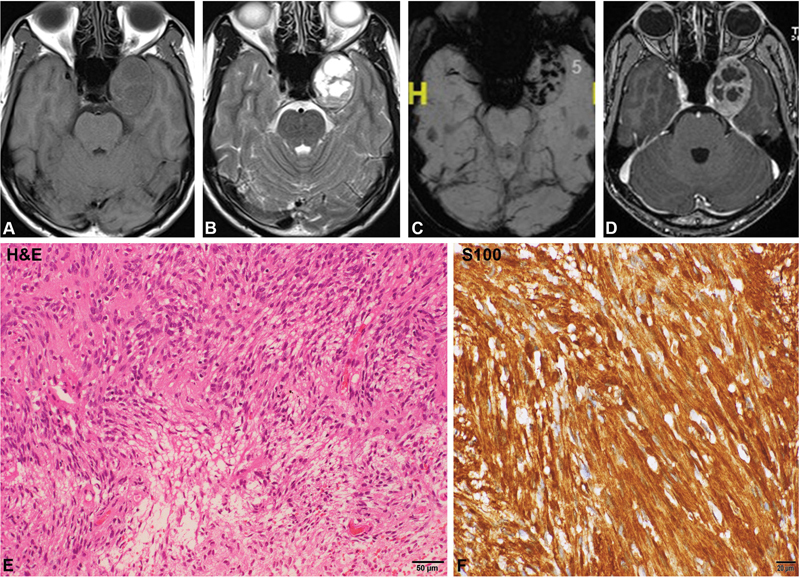
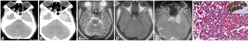
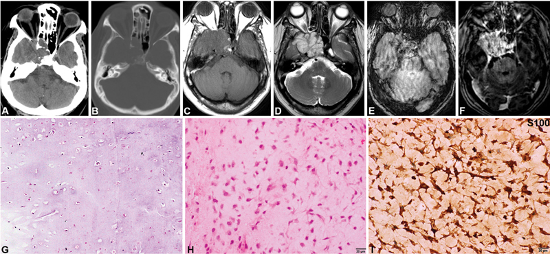
The cavernous sinus is a complex space composed of extradural venous plexus within dural folds. Several important structures like the carotid artery, cranial nerves, and sympathetic nerve fibers traverse through this space. Radiological diagnosis may not be definitive and in the context of discordance between clinical and neuroimaging diagnosis, histopathological evaluation becomes essential for diagnosis and management. Literature on the pathological spectrum of lesions is scarce as, with a shift in the treatment paradigm, most small lesions of cavernous sinus are treated with radiosurgery. However, surgical management still plays a role for larger lesions and in radiologically ambiguous cases for planning the definitive management. We retrospectively reviewed all surgically resected lesions of the cavernous sinus over the last two decades (1998-2019). The clinical presentation, neuroimaging features, and histopathological findings were reviewed. Lesions extending from sella and other adjacent areas were excluded. Thirty-eight cases of isolated cavernous sinus mass lesions were diagnosed over the last two decades (1998-2019). Cavernous hemangiomas (19 cases, 50%) constituted the most frequent pathology, followed by aspergilloma, meningioma, schwannoma, metastatic adenocarcinoma, chondrosarcoma, and chordoma. Overall, 29.4% (10/34) could not be accurately diagnosed on neuroimaging. Of these, four cases of cavernous hemangiomas were mistaken for either meningioma (three cases) or schwannoma (one case). Neither chordoma nor chondrosarcoma was suspected. This is the first study in literature, enumerating the pathological and imaging spectrum of surgically resected cavernous sinus lesions. Cavernous hemangiomas, metastases and chordomas, and chondrosarcoma posed the greatest difficulty in diagnosis on neuroimaging and the reasons for the same are analyzed. In the context of clinical and neuroimaging discordance in diagnosis, pathological characterization becomes essential for appropriate and timely management.
海绵窦是一个复杂的间隙,由硬脑膜皱襞内的硬膜外静脉丛组成。颈内动脉、脑神经和交感神经纤维等几个重要结构穿过该间隙。放射学诊断可能不明确,在临床与神经影像学诊断不一致的情况下,组织病理学评估对于诊断和治疗至关重要。由于治疗模式的转变,大多数海绵窦小病变采用放射外科治疗,关于病变病理谱的文献较少。然而,手术治疗对于较大病变以及在放射学表现不明确的病例中规划最终治疗仍发挥着作用。
我们回顾性分析了过去二十年(1998 - 2019年)所有经手术切除的海绵窦病变。对临床表现、神经影像学特征和组织病理学结果进行了回顾。排除了起源于鞍区和其他相邻区域的病变。
在过去二十年(1998 - 2019年)中,共诊断出38例孤立的海绵窦肿块病变。海绵状血管瘤(19例,占50%)是最常见的病理类型,其次是曲霉菌瘤、脑膜瘤、神经鞘瘤、转移性腺癌、软骨肉瘤和脊索瘤。总体而言,29.4%(10/34)的病例在神经影像学上无法准确诊断。其中,4例海绵状血管瘤被误诊为脑膜瘤(3例)或神经鞘瘤(1例)。未怀疑有脊索瘤或软骨肉瘤。
这是文献中第一项列举经手术切除的海绵窦病变的病理和影像学谱的研究。分析了海绵状血管瘤、转移瘤、脊索瘤和软骨肉瘤在神经影像学诊断中面临最大困难的原因。在临床和神经影像学诊断不一致的情况下,病理特征对于恰当及时的治疗至关重要。