Department of Radiology and Imaging Sciences, Indiana University School of Medicine, Indianapolis, Indiana, USA.
Krannert Cardiovascular Research Center, Indiana University School of Medicine/IU Health Cardiovascular Institute, Indianapolis, Indiana, USA.
J Appl Clin Med Phys. 2022 Oct;23(10):e13741. doi: 10.1002/acm2.13741. Epub 2022 Aug 11.
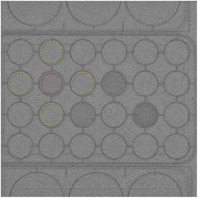
Interventional cardiology involves catheter-based treatment of heart disease, generally through fluoroscopically guided interventional procedures. Patients can be subject to considerable radiation dose due to prolonged fluoroscopy time and radiographic exposure, and therefore efforts to minimize patient dose should always be undertaken. Developing standardized, effective quality control programs for these systems is a difficult task owing to cross-vendor differences and automated control of imaging protocols. Furthermore, analyses of radiation dose should be performed in the context of its associated effects on image quality. The aim of the study is to investigate radiation dose and image quality in two fluoroscopic systems used for interventional cardiology procedures. Image quality was assessed in terms of spatial resolution and modulation transfer function, signal-to-noise and contrast-to-noise ratios, and spatial-temporal resolution of fluoroscopy and cineradiography images with phantoms simulating various patient thicknesses under routine cardiology protocols. The entrance air kerma (or air kerma rate) was measured and used to estimate entrance surface dose (or dose rate) in the phantoms.
介入心脏病学涉及基于导管的心脏病治疗,通常通过荧光镜引导的介入程序进行。由于荧光透视时间和放射暴露延长,患者可能会受到相当大的辐射剂量,因此应始终努力尽量减少患者剂量。由于跨供应商差异和成像协议的自动化控制,为这些系统制定标准化、有效的质量控制程序是一项艰巨的任务。此外,辐射剂量的分析应在其对图像质量的相关影响的背景下进行。本研究旨在调查用于介入心脏病学程序的两种荧光透视系统的辐射剂量和图像质量。使用模拟各种患者厚度的体模,根据空间分辨率和调制传递函数、信噪比和对比噪声比以及透视和电影摄影图像的空间-时间分辨率,评估图像质量。测量了入口空气比释动能(或空气比释动能率),并用于估计体模中的入口表面剂量(或剂量率)。