Rasul Sazan, Haug Alexander R
Department of Biomedical Imaging and Image-Guided Therapy, Division of Nuclear Medicine, Medical University of Vienna, 1090 Vienna, Austria.
Christian Doppler Laboratory for Applied Metabolomics (CDL AM), Medical University of Vienna, 1090 Vienna, Austria.
Cancers (Basel). 2022 Aug 2;14(15):3768. doi: 10.3390/cancers14153768.
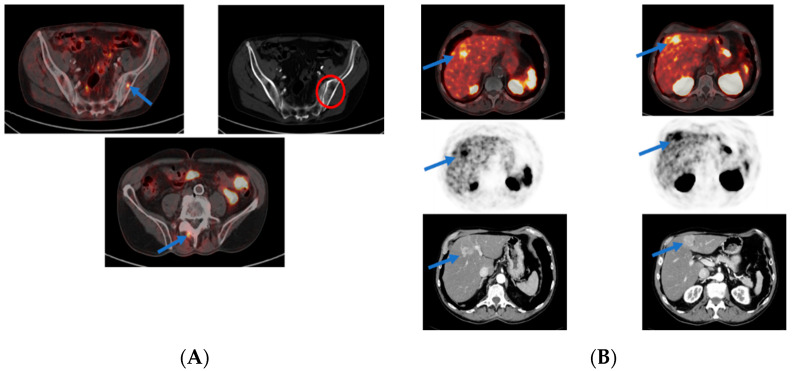
With the progressive aging of the population in industrially developed countries, as well as advances in diagnostic and biopsy techniques and improvements in patient awareness, the incidence of prostate cancer (PCa) is continuously increasing worldwide. Therefore, PCa is currently considered as the second leading cause of tumor-related death. Early detection of the tumor and its metastasis is essential, as the rate of disease recurrence is high and occurs in 27% to 53% of all patients who underwent curative therapy with radical prostatectomy or local radiotherapy. In this regard, the prostate specific membrane antigens, abbreviated as PSMAs, are type II membrane proteins that are highly expressed on the surface of malignant prostate tissue in PCa, particularly in aggressive, androgen-deprived, metastatic, and hormone-refractory PCa, and they are inversely associated with the androgen level. Up to 95% of adenocarcinomas of the prostate express PSMA receptors on their surface. Today, radionuclides that bind to these PSMA peptides are widely accepted for diagnostic and therapeutic purposes to specifically image and target prostate tumor cells at the molecular level, a process referred to as targeted theranostics. Numerous studies have demonstrated that the integration of these peptides into diagnostic and therapeutic procedures plays a critical role in the primary staging and treatment decisions of especially high-risk PCa, expands therapeutic options for patients with advanced stage of prostate tumor, and prolongs patients' survival rate. In this review article, we intend to briefly spotlight the latest clinical utilization of the PSMA-targeted radioligand PET imaging modality in patients with different stages of PCa. Furthermore, limitations and pitfalls of this diagnostic technique are presented.
随着工业发达国家人口的逐步老龄化,以及诊断和活检技术的进步以及患者意识的提高,前列腺癌(PCa)在全球范围内的发病率持续上升。因此,PCa目前被认为是肿瘤相关死亡的第二大主要原因。肿瘤及其转移的早期检测至关重要,因为疾病复发率很高,在所有接受根治性前列腺切除术或局部放疗的根治性治疗的患者中,有27%至53%会出现复发。在这方面,前列腺特异性膜抗原,简称为PSMAs,是II型膜蛋白,在PCa的恶性前列腺组织表面高度表达,特别是在侵袭性、雄激素剥夺性、转移性和激素难治性PCa中,并且它们与雄激素水平呈负相关。高达95%的前列腺腺癌在其表面表达PSMA受体。如今,与这些PSMA肽结合的放射性核素被广泛用于诊断和治疗目的,以在分子水平上特异性地成像和靶向前列腺肿瘤细胞,这一过程称为靶向诊疗。大量研究表明,将这些肽整合到诊断和治疗程序中,在尤其是高危PCa的初始分期和治疗决策中起着关键作用,扩大了前列腺肿瘤晚期患者的治疗选择,并延长了患者的生存率。在这篇综述文章中,我们打算简要介绍PSMA靶向放射性配体PET成像模式在不同阶段PCa患者中的最新临床应用。此外,还介绍了这种诊断技术的局限性和陷阱。