• 文献检索
  • 文档翻译
  • 深度研究
  • 学术资讯
  • Suppr Zotero 插件Zotero 插件
  • 邀请有礼
  • 套餐&价格
  • 历史记录
应用&插件
Suppr Zotero 插件Zotero 插件浏览器插件Mac 客户端Windows 客户端微信小程序
定价
高级版会员购买积分包购买API积分包
服务
文献检索文档翻译深度研究API 文档MCP 服务
关于我们
关于 Suppr公司介绍联系我们用户协议隐私条款
关注我们

Suppr 超能文献

核心技术专利:CN118964589B侵权必究
粤ICP备2023148730 号-1Suppr @ 2026

文献检索

告别复杂PubMed语法,用中文像聊天一样搜索,搜遍4000万医学文献。AI智能推荐,让科研检索更轻松。

立即免费搜索

文件翻译

保留排版,准确专业,支持PDF/Word/PPT等文件格式,支持 12+语言互译。

免费翻译文档

深度研究

AI帮你快速写综述,25分钟生成高质量综述,智能提取关键信息,辅助科研写作。

立即免费体验

静息状态下右颞叶癫痫患者全脑网络中的异常枢纽作为神经影像生物标志物

Abnormal hubs in global network as neuroimaging biomarker in right temporal lobe epilepsy at rest.

作者信息

Guo Ruimin, Zhao Yunfei, Jin Honghua, Jian Jihua, Wang Haibo, Jin Shengxi, Ren Hongwei

机构信息

Department of Medical Imaging, Tianyou Hospital of Wuhan University of Science and Technology, Wuhan, China.

Key Laboratory of Occupational Hazards and Identification, Wuhan University of Science and Technology, Wuhan, China.

出版信息

Front Psychiatry. 2022 Jul 27;13:981728. doi: 10.3389/fpsyt.2022.981728. eCollection 2022.

DOI:10.3389/fpsyt.2022.981728
PMID:35966487
原文链接:https://pmc.ncbi.nlm.nih.gov/articles/PMC9363580/
Abstract

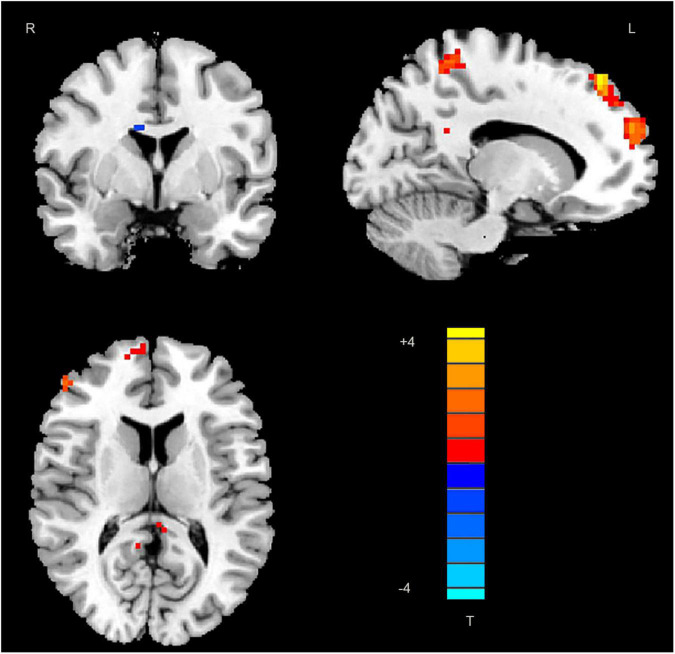
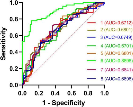
While abnormal neuroimaging features have been reported in patients suffering from right temporal lobe epilepsy (rTLE), the value of altered degree centrality (DC) as a diagnostic biomarker for rTLE has yet to be established. As such, the present study was designed to examine DC abnormalities in rTLE patients in order to gauge the diagnostic utility of these neuroimaging features. In total, 68 patients with rTLE and 73 healthy controls (HCs) participated in this study. Imaging data were analyzed using DC and receiver operating characteristic (ROC) methods. Ultimately, rTLE patients were found to exhibit reduced right caudate DC and increased left middle temporal gyrus, superior parietal gyrus, superior frontal gyrus, right precuneus, frontal gyrus Inferior gyrus, middle-superior frontal gyrus, and inferior parietal gyrus DC relative to HC. ROC analyses indicated that DC values in the right caudate nucleus could be used to differentiate between rTLE patients and HCs with a high degree of sensitivity and specificity. Together, these results thus suggest that rTLE is associated with abnormal DC values in the right caudate nucleus, underscoring the relevance of further studies of the underlying pathophysiology of this debilitating condition.

摘要

虽然已有报道称右侧颞叶癫痫(rTLE)患者存在异常神经影像学特征,但改变的度中心性(DC)作为rTLE诊断生物标志物的价值尚未确立。因此,本研究旨在检查rTLE患者的DC异常情况,以评估这些神经影像学特征的诊断效用。共有68例rTLE患者和73名健康对照者(HCs)参与了本研究。使用DC和受试者工作特征(ROC)方法对影像数据进行分析。最终发现,相对于HCs,rTLE患者右侧尾状核DC降低,左侧颞中回、顶上叶、额上回、右侧楔前叶、额下回、额中-上回及顶下叶DC增加。ROC分析表明,右侧尾状核的DC值可用于以高度敏感性和特异性区分rTLE患者和HCs。总之,这些结果表明rTLE与右侧尾状核DC值异常有关,强调了进一步研究这种使人衰弱病症潜在病理生理学的重要性。

https://cdn.ncbi.nlm.nih.gov/pmc/blobs/0ec6/9363580/684ea6586bf7/fpsyt-13-981728-g002.jpg
https://cdn.ncbi.nlm.nih.gov/pmc/blobs/0ec6/9363580/05243cd056ba/fpsyt-13-981728-g001.jpg
https://cdn.ncbi.nlm.nih.gov/pmc/blobs/0ec6/9363580/684ea6586bf7/fpsyt-13-981728-g002.jpg
https://cdn.ncbi.nlm.nih.gov/pmc/blobs/0ec6/9363580/05243cd056ba/fpsyt-13-981728-g001.jpg
https://cdn.ncbi.nlm.nih.gov/pmc/blobs/0ec6/9363580/684ea6586bf7/fpsyt-13-981728-g002.jpg

相似文献

1
Abnormal hubs in global network as neuroimaging biomarker in right temporal lobe epilepsy at rest.静息状态下右颞叶癫痫患者全脑网络中的异常枢纽作为神经影像生物标志物
Front Psychiatry. 2022 Jul 27;13:981728. doi: 10.3389/fpsyt.2022.981728. eCollection 2022.
2
Abnormal Degree Centrality as a Potential Imaging Biomarker for Right Temporal Lobe Epilepsy: A Resting-state Functional Magnetic Resonance Imaging Study and Support Vector Machine Analysis.异常度中心度作为右颞叶癫痫潜在的影像学生物标志物:一项静息态功能磁共振成像研究和支持向量机分析。
Neuroscience. 2022 Apr 1;487:198-206. doi: 10.1016/j.neuroscience.2022.02.004. Epub 2022 Feb 11.
3
The Bilateral Precuneus as a Potential Neuroimaging Biomarker for Right Temporal Lobe Epilepsy: A Support Vector Machine Analysis.双侧楔前叶作为右侧颞叶癫痫潜在的神经影像学生物标志物:支持向量机分析
Front Psychiatry. 2022 Jun 15;13:923583. doi: 10.3389/fpsyt.2022.923583. eCollection 2022.
4
Altered voxel-mirrored homotopic connectivity in right temporal lobe epilepsy as measured using resting-state fMRI and support vector machine analyses.使用静息态功能磁共振成像和支持向量机分析测量发现,右侧颞叶癫痫患者的体素镜像同伦连接发生改变。
Front Psychiatry. 2022 Jul 26;13:958294. doi: 10.3389/fpsyt.2022.958294. eCollection 2022.
5
Alteration of the alertness-related network in patients with right temporal lobe epilepsy: A resting state fMRI study.右侧颞叶癫痫患者警觉相关网络的改变:一项静息态功能磁共振成像研究。
Epilepsy Res. 2016 Nov;127:252-259. doi: 10.1016/j.eplepsyres.2016.09.013. Epub 2016 Sep 16.
6
Resting-state brain entropy in right temporal lobe epilepsy and its relationship with alertness.右颞叶癫痫的静息态脑熵及其与警觉性的关系。
Brain Behav. 2019 Nov;9(11):e01446. doi: 10.1002/brb3.1446. Epub 2019 Oct 12.
7
Altered brain network centrality in patients with adult comitant exotropia strabismus: A resting-state fMRI study.成人共同性外斜视患者脑网络中心性改变:一项静息态功能磁共振成像研究
J Int Med Res. 2018 Jan;46(1):392-402. doi: 10.1177/0300060517715340. Epub 2017 Jul 6.
8
Alteration of functional connectivity within visuospatial working memory-related brain network in patients with right temporal lobe epilepsy: a resting-state fMRI study.右侧颞叶癫痫患者视空间工作记忆相关脑网络功能连接的改变:一项静息态 fMRI 研究。
Epilepsy Behav. 2014 Jun;35:64-71. doi: 10.1016/j.yebeh.2014.04.001. Epub 2014 May 6.
9
Abnormal hubs in global network as potential neuroimaging marker in generalized anxiety disorder at rest.静息状态下全球网络中的异常枢纽作为广泛性焦虑症潜在的神经影像学标志物
Front Psychol. 2022 Dec 16;13:1075636. doi: 10.3389/fpsyg.2022.1075636. eCollection 2022.
10
Aberrant cerebral intrinsic activity and cerebro-cerebellar functional connectivity in right temporal lobe epilepsy: a resting-state functional MRI study.右侧颞叶癫痫患者脑内固有活动及脑-小脑功能连接异常:一项静息态功能磁共振研究。
Neuroreport. 2021 Aug 11;32(12):1009-1016. doi: 10.1097/WNR.0000000000001681.

引用本文的文献

1
Impaired degree centrality and effective connectivity contributed to deficits in cognition and depression in patients with temporal lobe epilepsy.颞叶癫痫患者的度中心性受损和有效连接性与认知功能障碍及抑郁有关。
Sci Rep. 2025 Jul 1;15(1):20466. doi: 10.1038/s41598-025-05347-9.
2
Altered Cerebro-Cerebellar Effective Connectivity in New-Onset Juvenile Myoclonic Epilepsy.新发青少年肌阵挛癫痫患者脑-小脑有效连接的改变
Brain Sci. 2022 Dec 3;12(12):1658. doi: 10.3390/brainsci12121658.

本文引用的文献

1
Abnormal Degree Centrality as a Potential Imaging Biomarker for Right Temporal Lobe Epilepsy: A Resting-state Functional Magnetic Resonance Imaging Study and Support Vector Machine Analysis.异常度中心度作为右颞叶癫痫潜在的影像学生物标志物:一项静息态功能磁共振成像研究和支持向量机分析。
Neuroscience. 2022 Apr 1;487:198-206. doi: 10.1016/j.neuroscience.2022.02.004. Epub 2022 Feb 11.
2
Temporal Lobe Epilepsy and Psychiatric Comorbidity.颞叶癫痫与精神共病
Front Neurol. 2021 Nov 30;12:775781. doi: 10.3389/fneur.2021.775781. eCollection 2021.
3
Aberrant Amplitude of Low-Frequency Fluctuation and Degree Centrality within the Default Mode Network in Patients with Vascular Mild Cognitive Impairment.
血管性轻度认知障碍患者默认模式网络内低频波动的异常幅度和中心度
Brain Sci. 2021 Nov 19;11(11):1534. doi: 10.3390/brainsci11111534.
4
Resting-state functional connectivity in the idiopathic generalized epilepsies: A systematic review and meta-analysis of EEG and MEG studies.特发性全身性癫痫的静息态功能连接性:脑电图和脑磁图研究的系统评价与荟萃分析
Epilepsy Behav. 2021 Oct 1;124:108336. doi: 10.1016/j.yebeh.2021.108336.
5
Thermal scanning probe lithography-a review.热扫描探针光刻技术综述
Microsyst Nanoeng. 2020 Apr 6;6:21. doi: 10.1038/s41378-019-0124-8. eCollection 2020.
6
Individual localization value of resting-state fMRI in epilepsy presurgical evaluation: A combined study with stereo-EEG.静息态 fMRI 在癫痫术前评估中的个体定位价值:与立体脑电图的联合研究。
Clin Neurophysiol. 2021 Dec;132(12):3197-3206. doi: 10.1016/j.clinph.2021.07.028. Epub 2021 Aug 30.
7
Network centrality for the identification of biomarkers in respondent-driven sampling datasets.基于网络中心度的应答驱动抽样数据集生物标志物识别。
PLoS One. 2021 Aug 24;16(8):e0256601. doi: 10.1371/journal.pone.0256601. eCollection 2021.
8
Graph-theory based degree centrality combined with machine learning algorithms can predict response to treatment with antiepileptic medications in children with epilepsy.基于图论的度数中心度结合机器学习算法可预测癫痫儿童对抗癫痫药物治疗的反应。
J Clin Neurosci. 2021 Sep;91:276-282. doi: 10.1016/j.jocn.2021.07.016. Epub 2021 Jul 26.
9
Characterization of Potential Protein Biomarkers for Major Depressive Disorder Using Matrix-Assisted Laser Desorption Ionization/Time-of-Flight Mass Spectrometry.使用基质辅助激光解吸电离/飞行时间质谱法对重度抑郁症的潜在蛋白质生物标志物进行表征。
Molecules. 2021 Jul 23;26(15):4457. doi: 10.3390/molecules26154457.
10
EEG p-adic quantum potential accurately identifies depression, schizophrenia and cognitive decline.脑电 p-adic 量子势准确识别抑郁、精神分裂症和认知能力下降。
PLoS One. 2021 Aug 5;16(8):e0255529. doi: 10.1371/journal.pone.0255529. eCollection 2021.