Department of Cardiac Surgery, Beijing Anzhen Hospital, Capital Medical University, Beijing, People's Republic of China.
Beijing Institute of Heart, Lung and Blood Vessel Diseases, Beijing, People's Republic of China.
Braz J Cardiovasc Surg. 2022 Aug 16;37(4):439-446. doi: 10.21470/1678-9741-2020-0587.
A weak venous wall is one of the major reasons contributing to vein graft failure after coronary artery bypass grafting (CABG). We investigated whether adventitial collagen cross-linking by glutaraldehyde reinforces venous wall, preserving the endothelium of veins during high-pressure distention.
Human saphenous veins (SVs) were collected from 40 patients undergoing CABG, and adventitia cross-linking was performed with 0.3% glutaraldehyde for five minutes. The cross-linked SVs were accessed by biodegradation assay, immunofluorescent staining, and tensile test. Native SVs and cross-linked SVs from another 20 patients received the 200 mmHg pressure distention for two minutes. Pressure-induced injury of SVs were accessed by immunohistochemistry and electron microscopy.
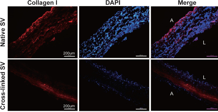
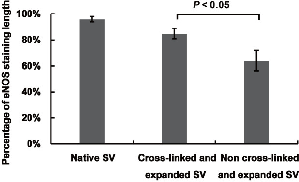
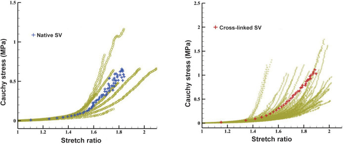
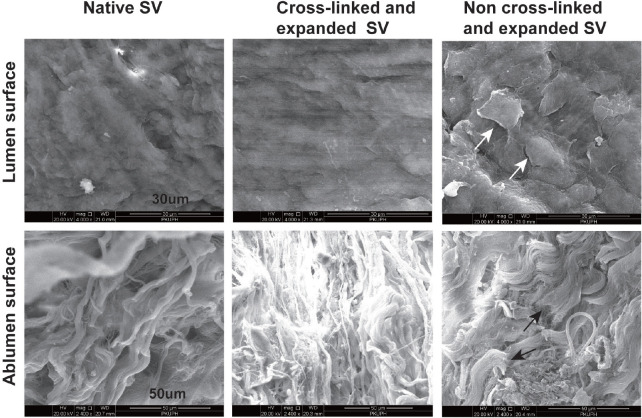
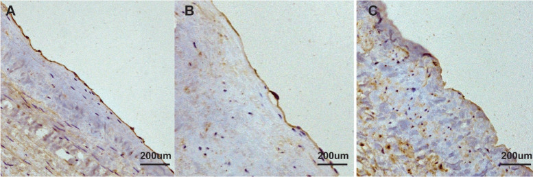
Time to digestion was 97±13 minutes for native SVs and 720±0 minutes for cross-linked SVs (P<0.05). After adventitial cross-linking, the collagen I fibres of the vein remarkably presented with compact and nonporous arrangement. In the high-stretch region (stretch ratio 1.4-1.8), the Young's elastic modulus of stress-stretch ratio curve in cross-linked SVs was larger than that in native SVs (13.88 vs. 5.83, P<0.05). The cross-linked SVs had a lower extent of endothelial denudation without fibre fracture during high-pressure distension than native SVs. Comparing with the non-cross-linked SVs, the percentage of endothelial nitric oxide synthase staining length on the endothelium of cross-linked SVs was significantly preserved after high-pressure distension (85.2% vs. 64.7%, P<0.05).
Adventitial collagen cross-linking by glutaraldehyde reinforced venous wall by increasing stiffness and decreasing extensibility of SVs and mitigated the endothelial damage under high-pressure distension.
静脉壁薄弱是冠状动脉旁路移植术后(CABG)静脉移植物失败的主要原因之一。我们研究了戊二醛交联血管外膜是否能在高压扩张时增强静脉壁,同时保持静脉内皮。
从 40 例行 CABG 的患者中采集人体大隐静脉(SV),用 0.3%戊二醛交联 5 分钟。通过生物降解试验、免疫荧光染色和拉伸试验评估交联的 SV。来自另外 20 名患者的天然 SV 和交联 SV 接受 200mmHg 的压力扩张 2 分钟。通过免疫组化和电子显微镜评估 SV 的压力损伤。
天然 SV 的消化时间为 97±13 分钟,交联 SV 为 720±0 分钟(P<0.05)。交联后,静脉胶原 I 纤维呈现致密且无孔的排列。在高拉伸区(拉伸比 1.4-1.8),交联 SV 的杨氏弹性模量与天然 SV 相比更大(13.88 比 5.83,P<0.05)。在高压扩张时,交联 SV 的内皮剥脱程度低于天然 SV,且没有纤维断裂。与未交联的 SV 相比,高压扩张后交联 SV 的内皮型一氧化氮合酶染色长度百分比显著保存(85.2%比 64.7%,P<0.05)。
戊二醛交联血管外膜通过增加 SV 的刚度和降低延展性来增强静脉壁,并减轻高压扩张下的内皮损伤。