Department of Neurology, Beth Israel Deaconess Medical Center, Harvard Medical School, Boston, Massachusetts, USA.
Department of Neurology, Massachusetts General Hospital, Harvard Medical School, Boston, Massachusetts, USA.
Stroke Vasc Neurol. 2023 Feb;8(1):26-33. doi: 10.1136/svn-2022-001653. Epub 2022 Aug 18.
We evaluate whether non-haemorrhagic imaging markers (NHIM) (white matter hyperintensity patterns, lacunes and enlarged perivascular spaces (EPVS)) can discriminate cerebral amyloid angiopathy (CAA) from hypertensive cerebral small vessel disease (HTN-cSVD) among patients with isolated lobar intracerebral haemorrhage (isolated-LICH).
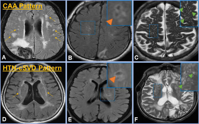
In patients with isolated-LICH, four cSVD aetiologic groups were created by incorporating the presence/distribution of NHIM: HTN-cSVD pattern, CAA pattern, mixed NHIM and no NHIM. CAA pattern consisted of patients with any combination of severe centrum semiovale EPVS, lobar lacunes or multiple subcortical spots pattern. HTN-cSVD pattern consisted of any HTN-cSVD markers: severe basal ganglia PVS, deep lacunes or peribasal ganglia white matter hyperintensity pattern. Mixed NHIM consisted of at least one imaging marker from either pattern. Our hypothesis was that patients with HTN-cSVD pattern/mixed NHIM would have a higher frequency of left ventricular hypertrophy (LVH), which is associated with HTN-cSVD.
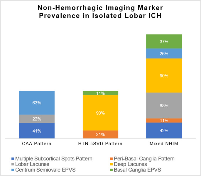
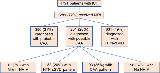
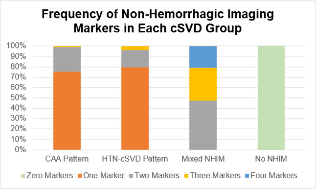
In 261 patients with isolated-LICH, CAA pattern was diagnosed in 93 patients, HTN-cSVD pattern in 53 patients, mixed NHIM in 19 patients and no NHIM in 96 patients. The frequency of LVH was similar among those with HTN-cSVD pattern and mixed NHIM (50% vs 39%, p=0.418) but was more frequent in HTN-cSVD pattern compared with CAA pattern (50% vs 20%, p<0.001). In a regression model, HTN-cSVD pattern (OR: 7.38; 95% CI 2.84 to 19.20) and mixed NHIM (OR: 4.45; 95% CI 1.25 to 15.90) were found to be independently associated with LVH.
Among patients with isolated-LICH, NHIM may help differentiate HTN-cSVD from CAA, using LVH as a marker for HTN-cSVD.
我们评估非出血性影像学标志物(NHIM)(脑白质高信号模式、腔隙和扩大的血管周围间隙(EPVS))是否可以在孤立性脑叶内出血(isolated-LICH)患者中区分脑淀粉样血管病(CAA)和高血压性脑小血管病(HTN-cSVD)。
在 isolated-LICH 患者中,通过合并 NHIM 的存在/分布,创建了四个 cSVD 病因学组:HTN-cSVD 模式、CAA 模式、混合 NHIM 和无 NHIM。CAA 模式由任何组合的严重半卵圆中心 EPVS、脑叶腔隙或多发性皮质下斑点模式的患者组成。HTN-cSVD 模式由任何 HTN-cSVD 标志物组成:严重基底节 PVS、深部腔隙或基底节周围脑白质高信号模式。混合 NHIM 由至少一个来自任何一种模式的影像学标志物组成。我们的假设是,HTN-cSVD 模式/混合 NHIM 的患者会有更高的左心室肥厚(LVH)频率,这与 HTN-cSVD 相关。
在 261 例 isolated-LICH 患者中,93 例诊断为 CAA 模式,53 例诊断为 HTN-cSVD 模式,19 例诊断为混合 NHIM,96 例诊断为无 NHIM。HTN-cSVD 模式和混合 NHIM 患者的 LVH 频率相似(50%与 39%,p=0.418),但 HTN-cSVD 模式较 CAA 模式更常见(50%与 20%,p<0.001)。在回归模型中,HTN-cSVD 模式(OR:7.38;95%CI 2.84 至 19.20)和混合 NHIM(OR:4.45;95%CI 1.25 至 15.90)被发现与 LVH 独立相关。
在 isolated-LICH 患者中,NHIM 可能有助于区分 HTN-cSVD 和 CAA,LVH 作为 HTN-cSVD 的标志物。