Farronato Marco, Farronato Davide, Giannì Aldo Bruno, Inchingolo Francesco, Nucci Ludovica, Tartaglia Gianluca Martino, Maspero Cinzia
Department of Orthodontics, Faculty of Medicine, University of Milan, 20142 Milano, Italy.
School of Medicine and Surgery, University of Insubria, Via G. Piatti 10, 21100 Varese, Italy.
Bioengineering (Basel). 2022 Aug 3;9(8):361. doi: 10.3390/bioengineering9080361.
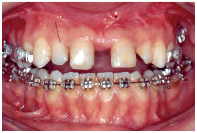
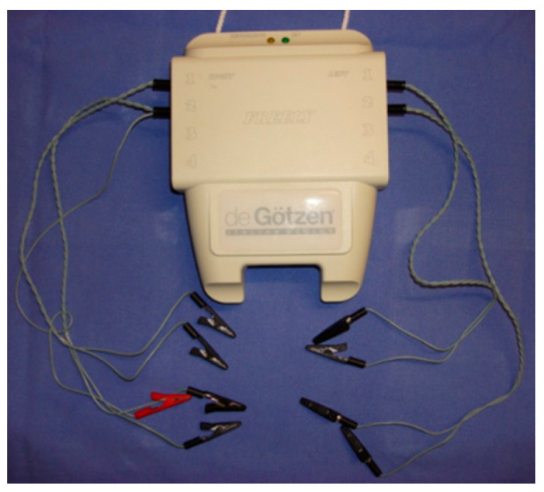
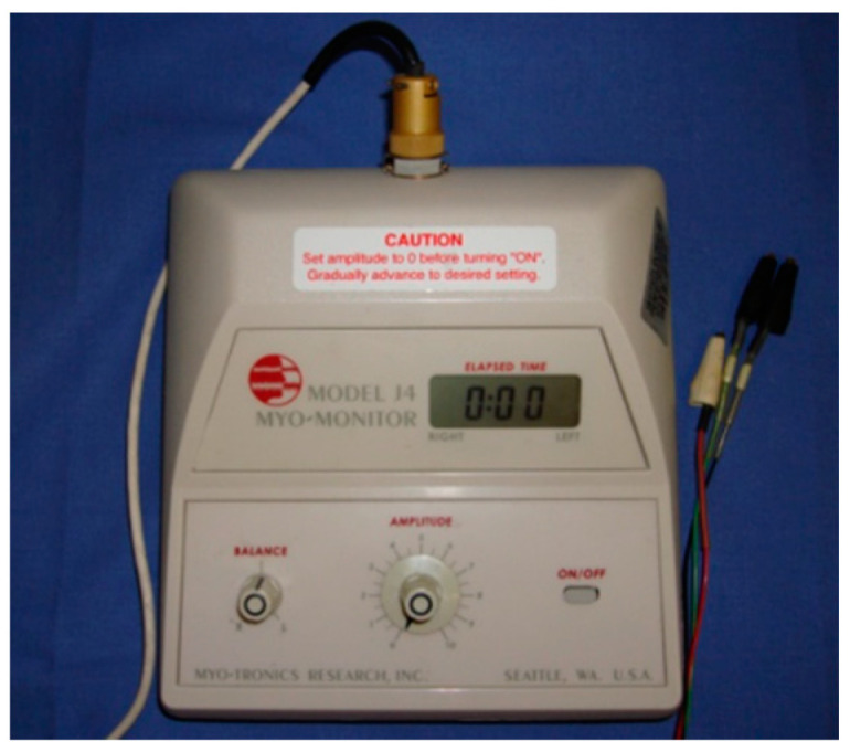
The study aims to investigate the modifications in the temporalis and the masseter activity in adult patients before and after SARPE (Surgically Assisted Rapid Palatal Expansion) by measuring electromyographic and electrokinesographic activity. 24 adult patients with unilateral posterior crossbite on the right side were selected from the Orthodontic Department of the University of Milan. Three electromyographic and electrokinesographic surface readings were taken respectively before surgery (T0) and 8 months after surgery (T1). The electromyographic data of both right and left masseter and anterior temporalis muscles were recorded during multiple tests: standardized maximum voluntary contraction (MVC)s, after transcutaneous electrical nerve stimulation (TENS) and at rest. T0 and T1 values were compared with paired Student’s t-test (p < 0.05). Results: Significant differences were found in the activity of right masseter (p = 0.03) and right temporalis (p = 0.02) during clench, in the evaluation of right masseter at rest (p = 0.03), also the muscular activity of masseters at rest after TENS from T0 to T1 (pr = 0.04, pl = 0.04). No significant differences were found in the activity of left masseter (p = 0.41) and left temporalis (p = 0.39) during clench and MVC, in the evaluation of left masseter at rest (p = 0.57) and in the activity during MVC of right masseter (p = 0.41), left masseter (p = 0.34), right temporalis (p = 0.51) and left temporalis (p = 0.77). Results showed that the activity of the masseter and temporalis muscles increased significantly after SARPE during rest and clenching on the side where the cross-bite was treated.
本研究旨在通过测量肌电图和运动电图活动,调查成年患者在外科辅助快速腭扩展术(SARPE)前后颞肌和咬肌活动的变化。从米兰大学正畸科选取了24例右侧单侧后牙反合的成年患者。在手术前(T0)和手术后8个月(T1)分别进行三次肌电图和运动电图表面读数。在多次测试中记录左右咬肌和颞肌前部的肌电图数据:标准化最大自主收缩(MVC)、经皮电神经刺激(TENS)后以及静息时。T0和T1值采用配对学生t检验进行比较(p < 0.05)。结果:在紧咬时,右侧咬肌(p = 0.03)和右侧颞肌(p = 0.02)的活动、静息时右侧咬肌的评估(p = 0.03)以及从T0到T1经TENS后咬肌静息时的肌肉活动(pr = 0.04,pl = 0.04)存在显著差异。在紧咬和MVC时,左侧咬肌(p = 0.41)和左侧颞肌(p = 0.39)的活动、静息时左侧咬肌的评估(p =