• 文献检索
  • 文档翻译
  • 深度研究
  • 学术资讯
  • Suppr Zotero 插件Zotero 插件
  • 邀请有礼
  • 套餐&价格
  • 历史记录
应用&插件
Suppr Zotero 插件Zotero 插件浏览器插件Mac 客户端Windows 客户端微信小程序
定价
高级版会员购买积分包购买API积分包
服务
文献检索文档翻译深度研究API 文档MCP 服务
关于我们
关于 Suppr公司介绍联系我们用户协议隐私条款
关注我们

Suppr 超能文献

核心技术专利:CN118964589B侵权必究
粤ICP备2023148730 号-1Suppr @ 2026

文献检索

告别复杂PubMed语法,用中文像聊天一样搜索,搜遍4000万医学文献。AI智能推荐,让科研检索更轻松。

立即免费搜索

文件翻译

保留排版,准确专业,支持PDF/Word/PPT等文件格式,支持 12+语言互译。

免费翻译文档

深度研究

AI帮你快速写综述,25分钟生成高质量综述,智能提取关键信息,辅助科研写作。

立即免费体验

柱状细胞病变是否是低级别乳腺肿瘤通路中最早的非必需前体?

Are Columnar Cell Lesions the Earliest Non-Obligate Precursor in the Low-Grade Breast Neoplasia Pathway?

机构信息

Department of Pathology and Laboratory Medicine, Faculty of Medicine, University of Ottawa, Ottawa, ON K1N 6N5, Canada.

Department of Pathology and Laboratory Medicine, Emory University Hospital, Emory University School of Medicine, Atlanta, GA 30322, USA.

出版信息

Curr Oncol. 2022 Aug 11;29(8):5664-5681. doi: 10.3390/curroncol29080447.

DOI:10.3390/curroncol29080447
PMID:36005185
原文链接:https://pmc.ncbi.nlm.nih.gov/articles/PMC9406596/
Abstract

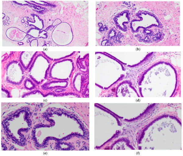
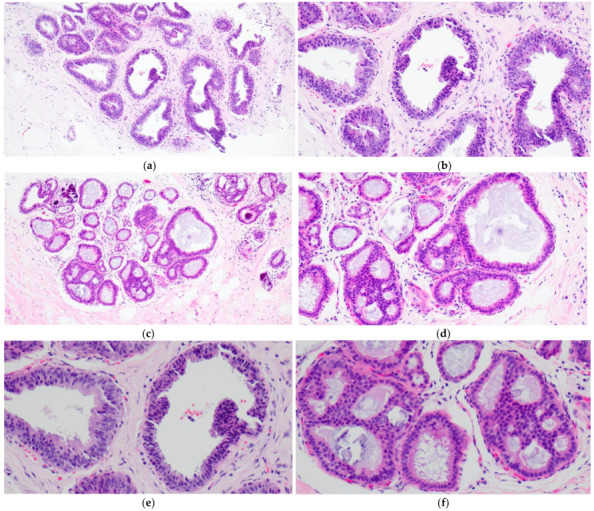
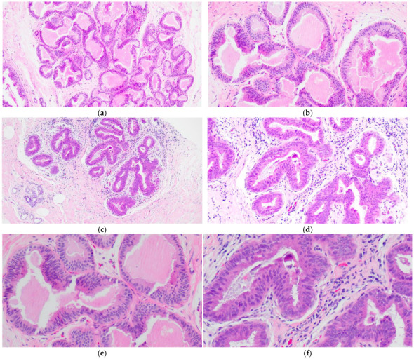
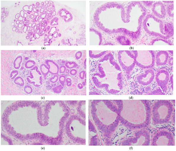
Columnar cell lesions (CCLs) of the breast comprise a spectrum of morphologic alterations of the terminal duct lobular unit involving variably dilated and enlarged acini lined by columnar epithelial cells. The World Health Organization currently classifies CCLs without atypia as columnar cell change (CCC) and columnar cell hyperplasia (CCH), whereas flat epithelial atypia (FEA) is a unifying term encompassing both CCC and CCH with cytologic atypia. CCLs have been increasingly recognized in stereotactic core needle biopsies (CNBs) performed for the assessment of calcifications. CCLs are believed to represent the earliest non-obligate precursor of low-grade invasive breast carcinomas as they share molecular alterations and often coexist with entities in the low-grade breast neoplasia pathway. Despite this association, however, the risk of progression of CCLs to invasive breast carcinoma appears low and may not exceed that of concurrent proliferative lesions. As the reported upgrade rates of pure CCL/FEA when identified as the most advanced high-risk lesion on CNB vary widely, the management of FEA diagnosed on CNB remains controversial. This review will include a historical overview of CCLs and will examine histologic diagnostic criteria, molecular alterations, prognosis and issues related to upgrade rates and clinical management.

摘要

乳腺柱状细胞病变(CCL)是一种累及终末导管小叶单位的形态学改变,包括各种程度的扩张和增大的腺泡,由柱状上皮细胞排列而成。目前,世界卫生组织将无异型的 CCL 分为柱状细胞改变(CCC)和柱状细胞增生(CCH),而扁平上皮异型性(FEA)是一个统一的术语,包括伴有细胞学异型性的 CCC 和 CCH。CCL 在用于评估钙化的立体定向核心针活检(CNB)中越来越被认识。CCL 被认为是低级别浸润性乳腺癌的最早非必需前体,因为它们具有分子改变,并且经常与低级别乳腺肿瘤途径中的实体共存。然而,尽管存在这种关联,但 CCL 进展为浸润性乳腺癌的风险似乎较低,并且可能不会超过同时存在的增生性病变的风险。由于在 CNB 上被确定为最高危病变时,纯 CCL/FEA 的报告升级率差异很大,因此在 CNB 上诊断出的 FEA 的管理仍然存在争议。这篇综述将包括 CCL 的历史概述,并将检查组织学诊断标准、分子改变、预后以及与升级率和临床管理相关的问题。

https://cdn.ncbi.nlm.nih.gov/pmc/blobs/f1f3/9406596/a8fb06cc74ac/curroncol-29-00447-g004.jpg
https://cdn.ncbi.nlm.nih.gov/pmc/blobs/f1f3/9406596/2d34b520cb67/curroncol-29-00447-g001.jpg
https://cdn.ncbi.nlm.nih.gov/pmc/blobs/f1f3/9406596/59fe0dd1c0a2/curroncol-29-00447-g002.jpg
https://cdn.ncbi.nlm.nih.gov/pmc/blobs/f1f3/9406596/d0f1749ef0ce/curroncol-29-00447-g003.jpg
https://cdn.ncbi.nlm.nih.gov/pmc/blobs/f1f3/9406596/a8fb06cc74ac/curroncol-29-00447-g004.jpg

相似文献

1
Are Columnar Cell Lesions the Earliest Non-Obligate Precursor in the Low-Grade Breast Neoplasia Pathway?柱状细胞病变是否是低级别乳腺肿瘤通路中最早的非必需前体?
Curr Oncol. 2022 Aug 11;29(8):5664-5681. doi: 10.3390/curroncol29080447.
2
Dimorphic cells: a common feature throughout the low nuclear grade breast neoplasia spectrum.两态细胞:低核级乳腺肿瘤谱中普遍存在的共同特征。
Virchows Arch. 2023 Feb;482(2):369-375. doi: 10.1007/s00428-022-03438-w. Epub 2022 Nov 15.
3
Proliferating activity in columnar cell lesions of the breast.乳腺柱状细胞病变中的增殖活性。
Virchows Arch. 2006 Dec;449(6):617-21. doi: 10.1007/s00428-006-0296-0. Epub 2006 Oct 6.
4
Are columnar cell lesions the earliest histologically detectable non-obligate precursor of breast cancer?柱状细胞病变是乳腺癌在组织学上可检测到的最早的非必然前驱病变吗?
Virchows Arch. 2008 Jun;452(6):589-98. doi: 10.1007/s00428-008-0609-6. Epub 2008 Apr 24.
5
Assessment of "grading" with Ki-67 and c-kit immunohistochemical expressions may be a helpful tool in management of patients with flat epithelial atypia (FEA) and columnar cell lesions (CCLs) on core breast biopsy.通过Ki-67和c-kit免疫组化表达评估“分级”可能是指导乳腺粗针活检中扁平上皮异型增生(FEA)和柱状细胞病变(CCL)患者管理的有用工具。
J Cell Physiol. 2009 Nov;221(2):343-9. doi: 10.1002/jcp.21858.
6
High frequency of coexistence of columnar cell lesions, lobular neoplasia, and low grade ductal carcinoma in situ with invasive tubular carcinoma and invasive lobular carcinoma.柱状细胞病变、小叶瘤变及低级别导管原位癌与浸润性小管癌和浸润性小叶癌共存的频率较高。
Am J Surg Pathol. 2007 Mar;31(3):417-26. doi: 10.1097/01.pas.0000213368.41251.b9.
7
Does flat epithelial atypia have rounder nuclei than columnar cell change/hyperplasia? A morphometric approach to columnar cell lesions of the breast.扁平上皮异型增生的细胞核比柱状细胞改变/增生的细胞核更圆吗?一种针对乳腺柱状细胞病变的形态测量方法。
Virchows Arch. 2016 Jun;468(6):663-73. doi: 10.1007/s00428-016-1923-z. Epub 2016 Mar 30.
8
Columnar cell lesions of the breast: an update and significance on core biopsy.乳腺柱状细胞病变:核心活检的最新进展及意义
Pathology. 2009 Jan;41(1):18-27. doi: 10.1080/00313020802563486.
9
Progression risk of columnar cell lesions of the breast diagnosed in core needle biopsies.在核心针活检中诊断的乳腺柱状细胞病变的进展风险。
Int J Cancer. 2011 Dec 1;129(11):2674-80. doi: 10.1002/ijc.25926. Epub 2011 Mar 25.
10
Non-operative breast pathology: columnar cell lesions.非手术性乳腺病理学:柱状细胞病变
J Clin Pathol. 2007 Dec;60(12):1307-12. doi: 10.1136/jcp.2006.040634. Epub 2006 Dec 20.

引用本文的文献

1
Canine, Feline, and Murine Mammary Tumors as a Model for Translational Research in Breast Cancer.犬、猫和鼠类乳腺肿瘤作为乳腺癌转化研究的模型
Vet Sci. 2025 Feb 19;12(2):189. doi: 10.3390/vetsci12020189.
2
[B3 lesions of the breast: histological, clinical, and epidemiological aspects : Update].[乳腺B3病变:组织学、临床及流行病学方面:最新进展]
Pathologie (Heidelb). 2023 Feb;44(1):5-16. doi: 10.1007/s00292-022-01180-3. Epub 2023 Jan 12.

本文引用的文献

1
Upgrade rate of percutaneously diagnosed pure flat epithelial atypia: systematic review and meta-analysis of 1,924 lesions.经皮诊断的单纯扁平上皮异型增生的升级率:对1924个病变的系统评价和荟萃分析
J Osteopath Med. 2022 Feb 14;122(5):253-262. doi: 10.1515/jom-2021-0206.
2
Nuclear morphology in breast lesions: refining its assessment to improve diagnostic concordance.乳腺病变中的核形态学:优化其评估以提高诊断一致性。
Histopathology. 2022 Feb;80(3):515-528. doi: 10.1111/his.14577. Epub 2021 Dec 2.
3
Blunt duct adenosis: a separate entity from columnar cell lesions?
钝性管腺病:与柱状细胞病变不同的实体?
J Clin Pathol. 2022 Jan;75(1):5-9. doi: 10.1136/jclinpath-2020-207359. Epub 2021 Apr 15.
4
Upgrade Rate of Pure Flat Epithelial Atypia Diagnosed at Core Needle Biopsy: A Systematic Review and Meta-Analysis.在核心针活检中诊断为单纯平坦上皮不典型性的升级率:系统评价和荟萃分析。
Radiol Imaging Cancer. 2021 Jan 22;3(1):e200116. doi: 10.1148/rycan.2021200116. eCollection 2021 Jan.
5
High-Risk Lesions Detected by MRI-Guided Core Biopsy: Upgrade Rates at Surgical Excision and Implications for Management.MRI 引导核心活检检测到的高危病变:手术切除时的升级率及对管理的影响。
AJR Am J Roentgenol. 2021 Mar;216(3):622-632. doi: 10.2214/AJR.20.23040. Epub 2021 Jan 13.
6
Is Excisional Biopsy Needed for Pure FEA Diagnosed on a Core Biopsy?对于在粗针活检中诊断出的纯FEA,是否需要进行切除活检?
Am Surg. 2020 Sep;86(9):1088-1090. doi: 10.1177/0003134820943546. Epub 2020 Aug 20.
7
The 2019 World Health Organization classification of tumours of the breast.《2019年世界卫生组织乳腺肿瘤分类》
Histopathology. 2020 Aug;77(2):181-185. doi: 10.1111/his.14091. Epub 2020 Jul 29.
8
High-risk lesions diagnosed at MRI-guided vacuum-assisted breast biopsy: imaging characteristics, outcome of surgical excision or imaging follow-up.MRI引导下真空辅助乳腺活检诊断出的高危病变:影像学特征、手术切除结果或影像学随访
Breast Cancer. 2020 May;27(3):405-414. doi: 10.1007/s12282-019-01032-8. Epub 2019 Dec 14.
9
Pure flat epithelial atypia identified on core needle biopsy does not require excision.在核心针活检中发现的单纯平坦上皮不典型增生不需要切除。
Eur J Surg Oncol. 2020 Feb;46(2):235-239. doi: 10.1016/j.ejso.2019.10.029. Epub 2019 Oct 24.
10
Flat Epithelial Atypia in Breast Core Needle Biopsies With Radiologic-Pathologic Concordance: Is Excision Necessary?乳腺芯针活检中与影像学-病理学一致的平坦上皮不典型性:是否需要切除?
Am J Surg Pathol. 2020 Feb;44(2):182-190. doi: 10.1097/PAS.0000000000001385.