Department of Breast Oncology, Juntendo University Faculty of Medicine, Tokyo, Japan.
Department of Human Pathology, Juntendo University Faculty of Medicine, Tokyo, Japan.
PLoS One. 2022 Aug 25;17(8):e0273513. doi: 10.1371/journal.pone.0273513. eCollection 2022.
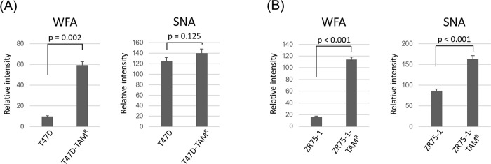
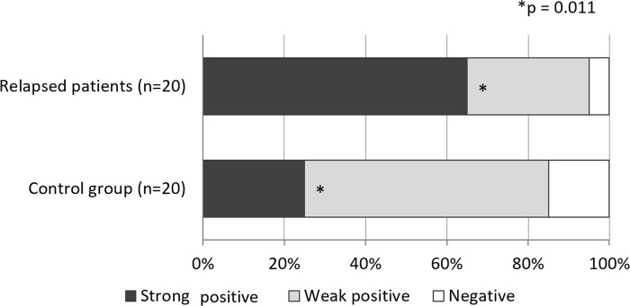
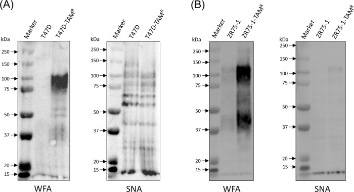
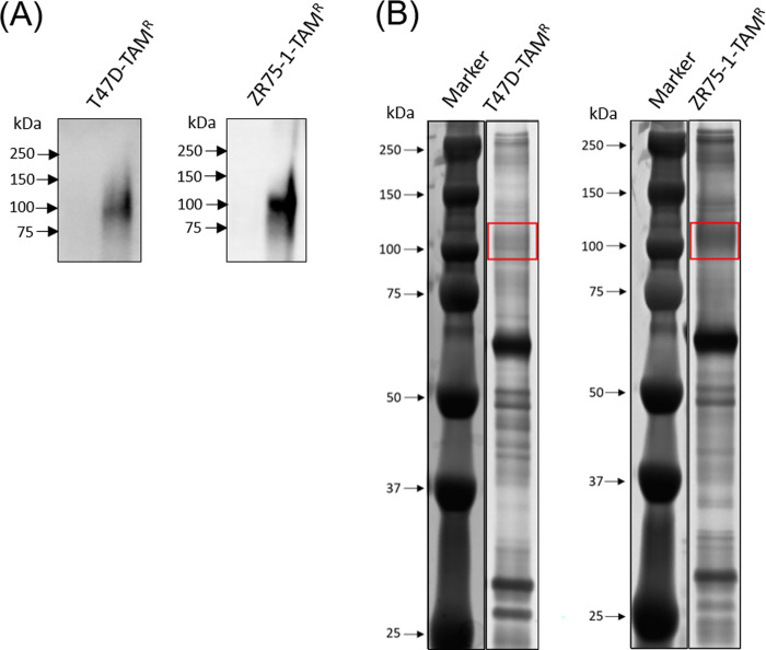
Glycosylation is one of the most important post-translational modifications of cell surface proteins involved in the proliferation, metastasis and treatment resistance of cancer cells. However, little is known about the role of glycosylation as the mechanism of breast cancer cell resistance to endocrine therapy. Herein, we aimed to identify the glycan profiles of tamoxifen-resistant human breast cancer cells, and their potential as predictive biomarkers for endocrine therapy. We established tamoxifen-resistant cells from estrogen receptor-positive human breast cancer cell lines, and their membrane-associated proteins were subjected to lectin microarray analysis. To confirm differential lectin binding to cellular glycoproteins, we performed lectin blotting analyses after electrophoretic separation of the glycoproteins. Mass spectrometry of the tryptic peptides of the lectin-bound glycoproteins was further conducted to identify glycoproteins binding to the above lectins. Finally, expression of the glycans that were recognized by a lectin was investigated using clinical samples from patients who received tamoxifen treatment after curative surgery. Lectin microarray analysis revealed that the membrane fractions of tamoxifen-resistant breast cancer cells showed increased binding to Wisteria floribunda agglutinin (WFA) compared to tamoxifen-sensitive cells. Glycoproteins seemed to be responsible for the differential WFA binding and the results of mass spectrometry revealed several membrane glycoproteins, such as CD166 and integrin beta-1, as candidates contributing to increased WFA binding. In clinical samples, strong WFA staining was more frequently observed in patients who had developed distant metastasis during tamoxifen treatment compared with non-relapsed patients. Therefore, glycans recognized by WFA are potentially useful as predictive markers to identify the tamoxifen-resistant and relapse-prone subset of estrogen receptor-positive breast cancer patients.
糖基化是参与癌细胞增殖、转移和治疗耐药的细胞表面蛋白最重要的翻译后修饰之一。然而,关于糖基化作为乳腺癌细胞对内分泌治疗耐药的机制知之甚少。在此,我们旨在鉴定他莫昔芬耐药的人乳腺癌细胞的聚糖图谱,及其作为内分泌治疗预测标志物的潜力。我们从雌激素受体阳性的人乳腺癌细胞系中建立了他莫昔芬耐药细胞,并对其膜相关蛋白进行了凝集素微阵列分析。为了确认差异结合细胞糖蛋白的凝集素,我们在糖蛋白电泳分离后进行了凝集素印迹分析。进一步对结合上述凝集素的糖蛋白的胰蛋白酶肽进行了质谱分析,以鉴定糖蛋白。最后,使用接受过治愈性手术后接受他莫昔芬治疗的患者的临床样本研究了被凝集素识别的聚糖的表达。凝集素微阵列分析显示,与他莫昔芬敏感细胞相比,他莫昔芬耐药乳腺癌细胞的膜部分与槐凝集素(WFA)的结合增加。糖蛋白似乎是导致差异 WFA 结合的原因,质谱结果表明几种膜糖蛋白,如 CD166 和整合素β-1,是导致 WFA 结合增加的候选物。在临床样本中,与未复发患者相比,在他莫昔芬治疗期间发生远处转移的患者中观察到强烈的 WFA 染色更为频繁。因此,WFA 识别的聚糖可能是一种有用的预测标志物,可用于识别雌激素受体阳性乳腺癌患者中他莫昔芬耐药和易复发的亚群。